Review
. 2020 Jan 14;4(4):478-492.
doi: 10.1002/hep4.1479. eCollection 2020 Apr.

Pathogenesis of Nonalcoholic Steatohepatitis: An Overview

Affiliations
Review

Pathogenesis of Nonalcoholic Steatohepatitis: An Overview

Gopanandan Parthasarathy et al. Hepatol Commun. .

Abstract

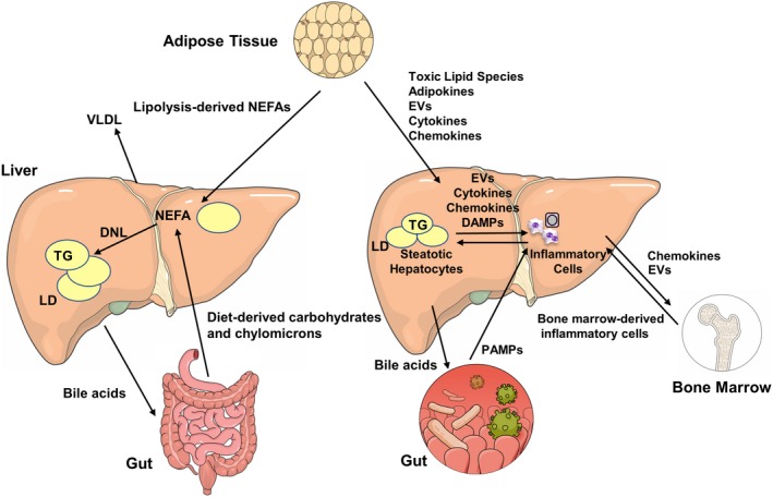
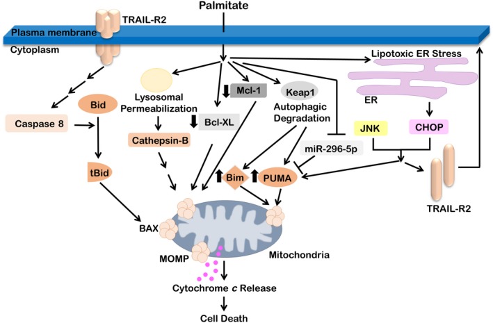
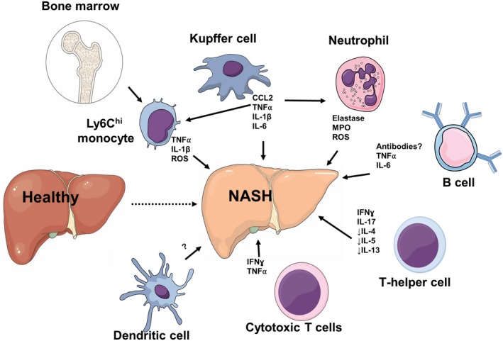
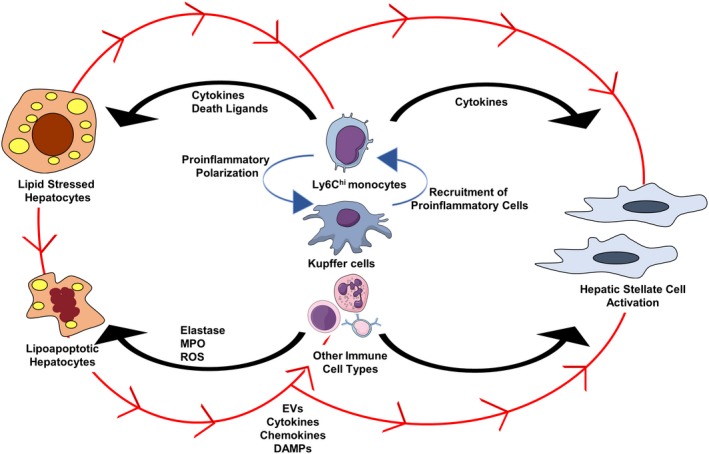
Nonalcoholic fatty liver disease (NAFLD) is a heterogeneous group of liver diseases characterized by the accumulation of fat in the liver. The heterogeneity of NAFLD is reflected in a clinical and histologic spectrum where some patients develop isolated steatosis of the liver, termed nonalcoholic fatty liver, whereas others develop hepatocyte injury, ballooning, inflammation, and consequent fibrosis, termed nonalcoholic steatohepatitis (NASH). Systemic insulin resistance is a major driver of hepatic steatosis in NAFLD. Lipotoxicity of accumulated lipids along with activation of the innate immune system are major drivers of NASH. Lipid-induced sublethal and lethal stress culminates in the activation of inflammatory processes, such as the release of proinflammatory extracellular vesicles and cell death. Innate and adaptive immune mechanisms involving macrophages, dendritic cells, and lymphocytes are central drivers of inflammation that recognize damage- and pathogen-associated molecular patterns and contribute to the progression of the inflammatory cascade. While the activation of the innate immune system and the recruitment of proinflammatory monocytes into the liver in NASH are well known, the exact signals that lead to this remain less well defined. Further, the contribution of other immune cell types, such as neutrophils and B cells, is an area of intense research. Many host factors, such as the microbiome and gut-liver axis, modify individual susceptibility to NASH. In this review, we discuss lipotoxicity, inflammation, and the contribution of interorgan crosstalk in NASH pathogenesis.


Figures

Figure 1
Figure 1
Metabolic interorgan crosstalk in NAFLD. This illustration depicts interorgan crosstalk in NAFL on the left and NASH on the right. Hepatic NEFAs are predominantly derived from three sources: lipolysis in adipose tissue, dietary lipid absorption, and DNL from carbohydrates in the liver. These NEFAs are stored in the liver as TG‐rich lipid droplets leading to hepatic steatosis or may be exported out of the liver as very low‐density lipoprotein to adipose tissue. Bile acids from the liver are key regulators of the gut–liver axis. Several mediators orchestrate the inflammatory milieu in the liver that results in NASH and fibrosis. Lipotoxic lipid species lead to hepatic stress and subsequent release of EVs, cytokines, chemokines, and DAMPs from liver cells. This results in recruitment of immune cells from the bone marrow. Bile acids from the liver, PAMPs from the gut, and adipokines from adipose tissues also influence various steps in this process. Abbreviations: LD, lipid droplet; VLDL, very low‐density lipoprotein.
Figure 2
Figure 2
Molecular pathways of palmitate‐induced lipotoxicity in hepatocytes. Palmitate activates the extrinsic death receptor‐mediated pathway of apoptosis and also activates the intrinsic pathway of apoptosis. Lysosomal permeabilization leads to the release of the protease cathepsin B. Lipotoxic ER stress leads to up‐regulation of the proapoptotic transcription factor CHOP. The stress‐induced kinase JNK and CHOP induce the death receptor TRAIL‐R2 and the proapoptotic Bcl‐2 family proteins PUMA and Bim. PUMA and Bim are also up‐regulated by palmitate‐induced autophagic degradation of Keap1. Palmitate decreases the expression of antiapoptotic proteins Mcl‐1 and Bcl‐XL. TRAIL‐R2 can undergo ligand‐independent oligomerization, cleavage‐induced activation of caspase 8, Bid cleavage to tBid, and activation of Bax. Ologomeric Bax results in mitochondrial outer membrane permeabilization, release of cytochrome c, activation of effector caspases, and apoptosis. Abbreviations: BAX, B‐cell lymphoma 2‐like protein 4; Bcl‐XL, B‐cell lymphoma‐extra large; Bim, B‐cell lymphoma 2‐like protein 11; Keap1, Kelch‐like ECH‐associated protein 1; Mcl‐1, induced myeloid leukemia cell differentiation protein; MOMP, major outer membrane protein; tBid, truncated p15 BID.
Figure 3
Figure 3
Immune dysregulation in the pathogenesis of NASH. Activation of liver‐resident KCs results in the release of CCL2 and other proinflammatory cytokines, such as TNFα, IL‐1, and IL‐6, leading to the recruitment of bone marrow‐derived Ly6Chi monocytes and neutrophils that further contribute to the inflammatory response. Activated neutrophils promote NASH by releasing elastase, MPO, and ROS. B cells can accumulate during NASH and produce TNFα and IL‐6. NASH is characterized by excessive Th1‐ and Th17‐derived IFNγ and IL‐17, respectively, and a deficiency in Th2‐derived IL‐4, IL‐5, and IL‐13. Cytotoxic CD8+ T cells are supported by type I IFN responses and lead to the production of IFNγ and TNFα. The role of DCs and their subsets in NASH is unclear as animal studies show contradictory results and it has not been investigated in humans. Abbreviation: ROS, reactive oxygen species.
Figure 4
Figure 4
Triadic lesion in the pathogenesis of NASH. Hepatocyte injury, macrophage‐mediated inflammation, and hepatic stellate cell activation comprise the key mechanistic abnormalities in NASH. Soluble and EV signals from hepatocytes lead to proinflammatory activation of macrophages, can recruit proinflammatory monocytes into the liver, and also lead to hepatic stellate cell activation (depicted in red arrows). Other immune cells, such as neutrophils and B cells, may also respond to hepatocyte‐derived signals. Activated macrophages release cytokines and chemokines that can promote hepatocyte apoptosis, attract other immune cells into the liver, and also influence the activation of hepatic stellate cells. Abbreviation: ROS, reactive oxygen species.

References

    1. Younossi ZM, Blissett D, Blissett R, Henry L, Stepanova M, Younossi Y, et al. The economic and clinical burden of nonalcoholic fatty liver disease in the United States and Europe. Hepatology 2016;64:1577‐1586. - PubMed
    1. Friedman SL, Neuschwander‐Tetri BA, Rinella M, Sanyal AJ. Mechanisms of NAFLD development and therapeutic strategies. Nat Med 2018;24:908‐922. - PMC - PubMed
    1. Kleiner DE, Brunt EM, Van Natta M, Behling C, Contos MJ, Cummings OW, et al.; Nonalcoholic Steatohepatitis Clinical Research Network . Design and validation of a histological scoring system for nonalcoholic fatty liver disease. Hepatology 2005;41:1313‐1321. - PubMed
    1. Hagstrom H, Nasr P, Ekstedt M, Hammar U, Stal P, Hultcrantz R, et al. Fibrosis stage but not NASH predicts mortality and time to development of severe liver disease in biopsy‐proven NAFLD. J Hepatol 2017;67:1265‐1273. - PubMed
    1. Hoang SA, Oseini A, Feaver RE, Cole BK, Asgharpour A, Vincent R, et al. Gene expression predicts histological severity and reveals distinct molecular profiles of nonalcoholic fatty liver disease. Sci Rep 2019;9:12541. - PMC - PubMed