Review
. 2019 Oct 29;3(1):11-20.
doi: 10.1021/acsptsci.9b00064. eCollection 2020 Feb 14.

The Role of Bone Morphogenetic Proteins in Diabetic Complications

Affiliations
Review

The Role of Bone Morphogenetic Proteins in Diabetic Complications

Nimna Perera et al. ACS Pharmacol Transl Sci. .

Abstract

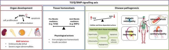
The prevalence of diabetes has reached epidemic proportions and is placing a significant burden on healthcare systems globally. Diabetes has a detrimental impact on many organs in the human body, including accelerating the development of micro- and macrovascular complications. Current therapeutic options to treat diabetic complications have their limitations. Importantly, many slow but fail to reverse the progression of diabetic complications. Bone morphogenetic proteins (BMPs) are a highly conserved subgroup of the transforming growth factor β (TGFβ) superfamily, signaling via serine/threonine kinase receptors, that have recently been implicated in glucose homeostasis and insulin resistance in the setting of diabetes. Downstream of the receptors, the signal can be transduced via the canonical Smad-dependent pathway or the noncanonical Smad-independent pathways. BMPs are essential in organ development, tissue homeostasis, and, as expected, disease pathogenesis. In fact, deletion of BMPs can be embryonically lethal or result in severe organ abnormalities. This review outlines the BMP signaling pathway and its relevance to diabetic complications, namely, diabetic nephropathy, diabetes-associated cardiovascular diseases, and diabetic retinopathy. Understanding the complexities of BMP signaling and particularly its tissue-, cellular-, and time-dependent actions will help delineate the underlying pathogenesis of the disease and may ultimately be harnessed in the treatment of diabetes-induced complications. This would replicate progress made in numerous other diseases, including cancer and atherosclerosis.


Conflict of interest statement

The authors declare no competing financial interest.

Figures

Figure 1
Figure 1
TGFβ and BMPs signaling pathways. (A) In the canonical pathway, TGFβ and BMPs bind to their respective receptors resulting in phosphorylation of Smad2/3 or Smad1/5/9, respectively. Common Smad4 binds to both Smad complexes and facilitates translocation to the nucleus, thereby activating transcription. (B) Alternatively, after receptor binding, the signal can be transduced via several noncanonical pathways including nitogen-activated protein kinase (MAPK), phosphoinositide 3-kinase (PI3K), and c-Jun N-terminal kinase (JNK), also resulting in gene transcription. Akt, protein kinase B; CRE, cyclic adenosine-monophosphate response element; ERK, extracellular signal-regulated kinases; MEK, mitogen-activated protein kinase kinase; mTOR, mammalian target of rapamycin; SBE, Smad binding element; TAK1, TGFβ-activated kinase 1; TAB1, TAK-1 binding protein 1. Figure created using artwork provided by Somesault1824, licensed under a Creative Commons License (CC BY-NC-SA 4.0).
Figure 2
Figure 2
Overview of BMPs actions in organ development, tissue homeostasis, and disease pathology. BMPs play a role in several cellular processes important in organ development, with BMP deletion leading to severe organ abnormalities. Beyond development, the TGFβ/BMP signaling is finely balanced, maintaining tissue homeostasis. When this balance is lost, aberrant signaling drives tissue remodelling in several organs, including organs commonly affected by diabetes. Figure created using artwork provided by Servier Medical Art by Servier, licensed under a Creative Commons License (CC BY 3.0).

References

    1. Ogurtsova K.; da Rocha Fernandes J. D.; Huang Y.; Linnenkamp U.; Guariguata L.; Cho N. H.; Cavan D.; Shaw J. E.; Makaroff L. E. (2017) IDF Diabetes Atlas: Global estimates for the prevalence of diabetes for 2015 and 2040. Diabetes Res. Clin. Pract. 128, 40–50. 10.1016/j.diabres.2017.03.024. - DOI - PubMed
    1. Adler A. I.; Boyko E. J.; Ahroni J. H.; Stensel V.; Forsberg R. C.; Smith D. G. (1997) Risk factors for diabetic peripheral sensory neuropathy. Results of the Seattle Prospective Diabetic Foot Study. Diabetes Care 20 (7), 1162–7. 10.2337/diacare.20.7.1162. - DOI - PubMed
    1. Semeraro F.; Cancarini A.; Dell’Omo R.; Rezzola S.; Romano M. R.; Costagliola C. (2015) Diabetic Retinopathy: Vascular and Inflammatory Disease. J. Diabetes Res. 2015, 582060.10.1155/2015/582060. - DOI - PMC - PubMed
    1. Katagiri T.; Watabe T. (2016) Bone Morphogenetic Proteins. Cold Spring Harbor Perspect. Biol. 8 (6), a021899.10.1101/cshperspect.a021899. - DOI - PMC - PubMed
    1. Urist M. R. (1965) Bone: formation by autoinduction. Science 150 (3698), 893–9. 10.1126/science.150.3698.893. - DOI - PubMed

LinkOut - more resources