. 2020 Jan 23;3(1):76-87.
doi: 10.1021/acsptsci.9b00095. eCollection 2020 Feb 14.

Serelaxin and the AT2 Receptor Agonist CGP42112 Evoked a Similar, Nonadditive, Cardiac Antifibrotic Effect in High Salt-Fed Mice That Were Refractory to Candesartan Cilexetil

Affiliations

Serelaxin and the AT2 Receptor Agonist CGP42112 Evoked a Similar, Nonadditive, Cardiac Antifibrotic Effect in High Salt-Fed Mice That Were Refractory to Candesartan Cilexetil

Yan Wang et al. ACS Pharmacol Transl Sci. .

Abstract

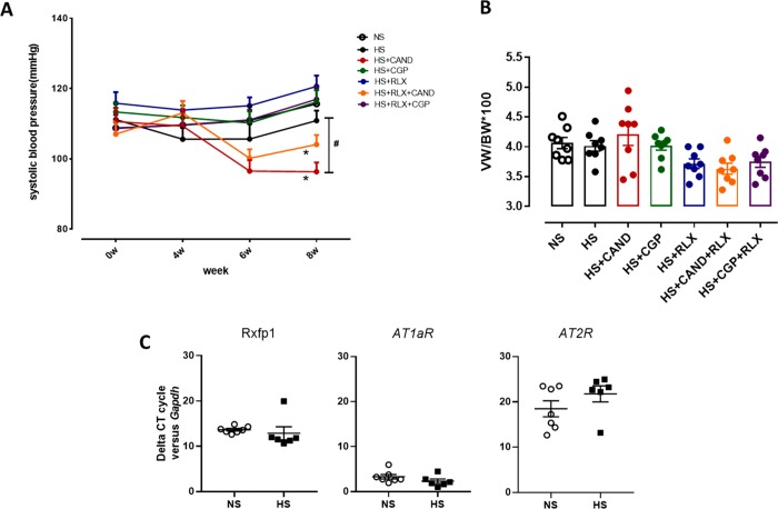
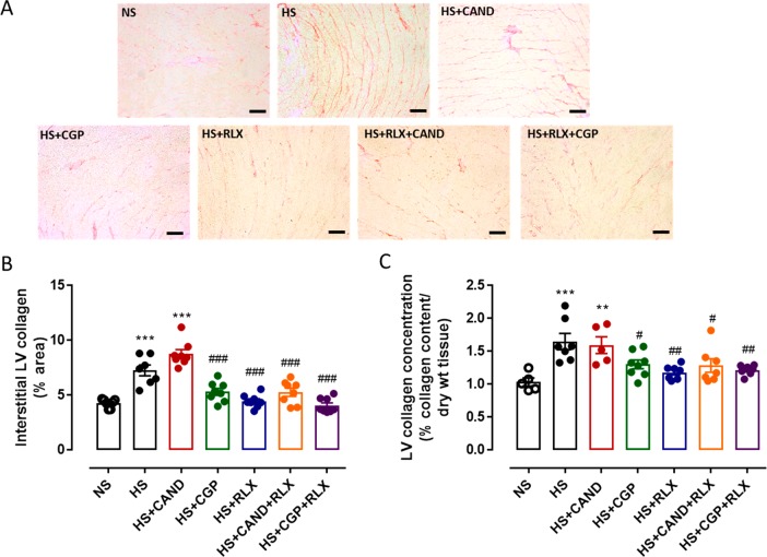
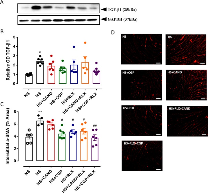
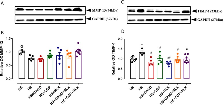
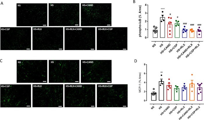
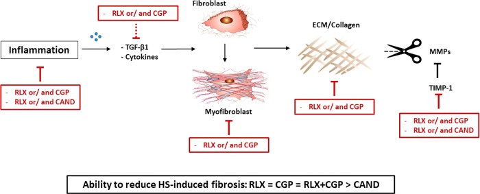
Fibrosis is involved in the majority of cardiovascular diseases and is a key contributor to end-organ dysfunction. In the current study, the antifibrotic effects of recombinant human relaxin-2 (serelaxin; RLX) and/or the AT2R agonist CGP42112 (CGP) were compared with those of the established AT1R antagonist, candesartan cilexetil (CAND), in a high salt-induced cardiac fibrosis model. High salt (HS; 5%) for 8 weeks did not increase systolic blood pressure in male FVB/N mice, but CAND treatment alone significantly reduced systolic blood pressure from HS-induced levels. HS significantly increased cardiac interstitial fibrosis, which was reduced by either RLX and/or CGP, which were not additive under the current experimental conditions, while CAND failed to reduce HS-induced cardiac fibrosis. The antifibrotic effects induced by RLX and/or CGP were associated with reduced myofibroblast differentiation. Additionally, all treatments inhibited the HS-induced elevation in tissue inhibitor of matrix metalloproteinases-1, together with trends for increased MMP-13 expression, that collectively would favor collagen degradation. Furthermore, these antifibrotic effects were associated with reduced cardiac inflammation. Collectively, these results highlight that either RXFP1 or AT2R stimulation represents novel therapeutic strategies to target fibrotic conditions, particularly in HS states that may be refractory to AT1R blockade.


Conflict of interest statement

The authors declare no competing financial interest.

Figures

Figure 1
Figure 1
Effects of RLX (0.5 mg/kg/day), CGP (1.44 mg/kg/day), CAND (2 mg/kg/day) or RLX with CAND or CGP on (A) systolic blood pressure measured by tail cuff and (B) ventricular weight (VW) to body weight (BW) ratio in FVB/N mice fed a high salt (HS, 5% NaCl) diet for 8 weeks. (C) Left ventricular expression of RXFP1, AT1aR, and AT2R mRNA levels from NS- and HS-treated mice; shown in the comparative cycle of threshold fluorescence at which RXFP1, AT1aR, and AT2R mRNA expression was detected, relative to the internal housekeeping gene GAPDH, by real-time PCR analysis. All treatments were given for 4 weeks, between weeks 5–8 of the HS diet. At the same time, a group of mice were fed a normal salt (NS, 0.5% NaCl) diet for comparison. All data are expressed as mean ± s.e.m. (n = 6–8 per group): (#) P < 0.05 vs HS for systolic blood pressure at 8 weeks (one-way ANOVA with Tukey correction for multiple comparisons); (∗) P < 0.05 for systolic blood pressure-time interaction compared with HS group (2-way repeated measures ANOVA).
Figure 2
Figure 2
(A) Representative images for interstitial fibrosis stained with picrosirius red (indicated by red) in left ventricle (LV) sections obtained from FVB/N mice fed on a normal salt (NS, 0.5% NaCl) diet or mice fed on a high salt (HS, 5% NaCl) diet in the presence or absence of RLX (0.5 mg/kg/day), CGP (1.44 mg/kg/day), CAND (2 mg/kg/day), or RLX with CAND or CGP. Scale bar for all images = 50 μm. (B) Group data of % area of interstitial fibrosis in LV sections, or (C) total tissue collagen measured by hydroxyproline assay in LV tissue from same animal groups in which picrosirius red staining was performed (n = 7–8 per group). All data are expressed as mean ± s.e.m. (∗∗) P < 0.01, (∗∗∗) P < 0.001 vs NS; (#) P < 0.05, (##) P < 0.01, (###) P < 0.001 vs HS, determined by One-Way ANOVA with Tukey correction for multiple comparisons.
Figure 3
Figure 3
(A) Representative Western blots of TGF-β1 (25 kDa) and GAPDH (37 kDa) from LV obtained from FVB/N mice fed on a normal salt (NS, 0.5% NaCl) diet or mice fed on a high salt (HS, 5% NaCl) diet in the presence or absence of RLX (0.5 mg/kg/day), CGP (1.44 mg/kg/day), CAND (2 mg/kg/day), or RLX with CAND or CGP (n = 5–7 per group). (B) Densitometric quantification of Western blots of TGF-β1 protein expression from indicated groups. Loading was adjusted by normalizing to GAPDH protein expression for each animal. All treatments were presented as a ratio to NS. (C) α-SMA expression quantified as % area from indicated groups. (n = 4–6 per group). All data are expressed as mean ± s.e.m. (∗) P < 0.05, (∗∗) P < 0.01 vs NS; (#) P < 0.05, (##) P < 0.01 vs HS, determined by One-Way ANOVA with Tukey correction for multiple comparisons. (D) Representative immunofluorescence staining images of α-SMA from same treatment groups as panel C. Scale bar for all images = 50 μm.
Figure 4
Figure 4
Representative Western blots of (A) MMP-13 (54 kDa) and (C) TIMP-1 (23 kDa) from LV obtained from FVB/N mice fed on a normal salt (NS, 0.5% NaCl) diet or mice fed on a high salt (HS, 5% NaCl) diet in the presence or absence of RLX (0.5 mg/kg/day), CGP (1.44 mg/kg/day), CAND (2 mg/kg/day), or RLX with CAND or CGP (n = 4–6 per group). Densitometric quantification of (B) MMP-13 and (D) TIMP-1 protein expressions from indicated groups. Protein loading was adjusted by normalizing to GAPDH protein expression for each animal. All treatments were presented as a ratio to NS. All data are expressed as mean ± s.e.m. (∗) P < 0.05 vs NS; (#) P < 0.05, (##) P < 0.01 vs HS, determined by One-Way ANOVA with Tukey correction for multiple comparisons.
Figure 5
Figure 5
Representative images for (A) p-IκB and (C) MCP-1 in LV sections obtained from FVB/N mice fed on a normal salt (NS, 0.5% NaCl) diet or mice fed on a high salt (HS, 5% NaCl) diet in the presence or absence of RLX (0.5 mg/kg/day), CGP (1.44 mg/kg/day), CAND (2 mg/kg/day) or RLX with CAND or CGP. Scale bar for all images = 50 μm. Group data of % area of (B) p-IκB and (D) MCP-1 in LV sections from indicated groups (n = 4–6 per group). All data are expressed as mean ± s.e.m. (∗∗) P < 0.01, (∗∗∗) P < 0.001 vs NS; (#) P < 0.05, (###) P < 0.001 vs HS, determined by One-Way ANOVA with Tukey correction for multiple comparisons.
Figure 6
Figure 6
Potential mechanisms involved in the antifibrotic actions of RLX and CGP, with inhibitory mechanisms depicted by red line blocks. RXFP1 and AT2R stimulation reduced inflammatory and pro-fibrotic factors thereby inhibiting myofibroblast differentiation and ECM production while enabling ECM degradation by inhibiting TIMP-1. In contrast, CAND inhibited fewer mechanisms in this pathway. Line blocks represent inhibition; dotted line block represents inhibitory trend.

References

    1. Kong P.; Christia P.; Frangogiannis N. G. (2014) The pathogenesis of cardiac fibrosis. Cell. Mol. Life Sci. 71 (4), 549–74. 10.1007/s00018-013-1349-6. - DOI - PMC - PubMed
    1. Zhou D.; Liu Y. (2016) Renal fibrosis in 2015: Understanding the mechanisms of kidney fibrosis. Nat. Rev. Nephrol. 12 (2), 68–70. 10.1038/nrneph.2015.215. - DOI - PMC - PubMed
    1. Lv W.; Booz G. W.; Wang Y.; Fan F.; Roman R. J. (2018) Inflammation and renal fibrosis: Recent developments on key signaling molecules as potential therapeutic targets. Eur. J. Pharmacol. 820, 65–76. 10.1016/j.ejphar.2017.12.016. - DOI - PMC - PubMed
    1. Prabhu S. D.; Frangogiannis N. G. (2016) The Biological Basis for Cardiac Repair After Myocardial Infarction: From Inflammation to Fibrosis. Circ. Res. 119 (1), 91–112. 10.1161/CIRCRESAHA.116.303577. - DOI - PMC - PubMed
    1. Wynn T. A.; Vannella K. M. (2016) Macrophages in Tissue Repair, Regeneration, and Fibrosis. Immunity 44 (3), 450–62. 10.1016/j.immuni.2016.02.015. - DOI - PMC - PubMed

LinkOut - more resources