. 2019 Nov 25;3(1):119-134.
doi: 10.1021/acsptsci.9b00079. eCollection 2020 Feb 14.

Venom Peptides with Dual Modulatory Activity on the Voltage-Gated Sodium Channel NaV1.1 Provide Novel Leads for Development of Antiepileptic Drugs

Affiliations

Venom Peptides with Dual Modulatory Activity on the Voltage-Gated Sodium Channel NaV1.1 Provide Novel Leads for Development of Antiepileptic Drugs

Chun Yuen Chow et al. ACS Pharmacol Transl Sci. .

Abstract

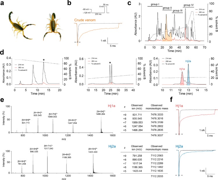
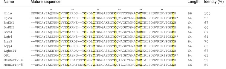
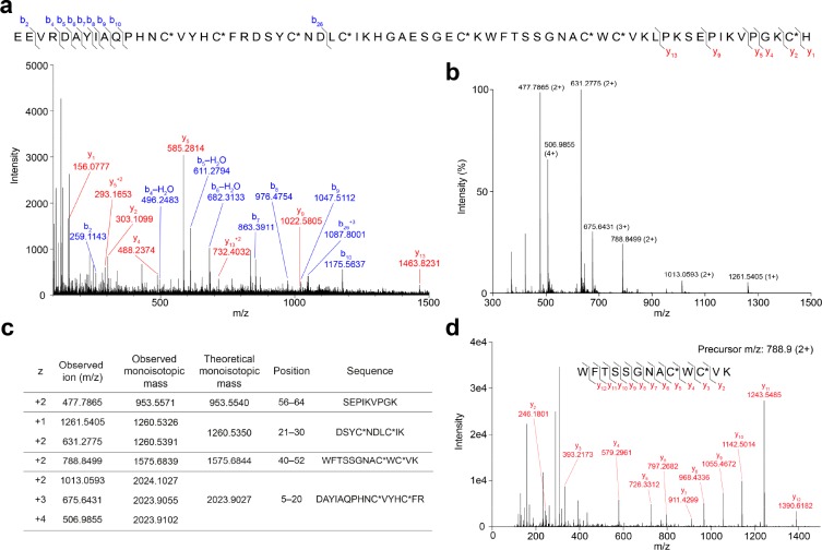
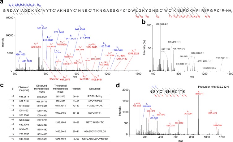
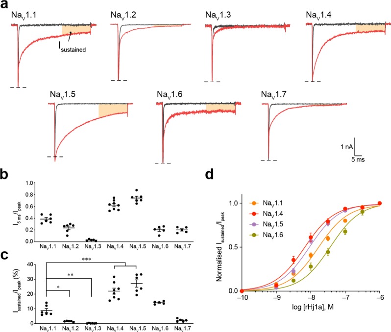
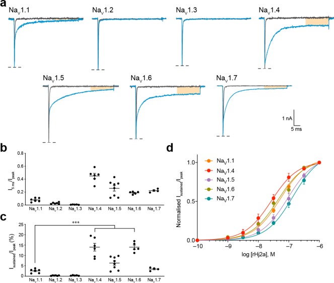
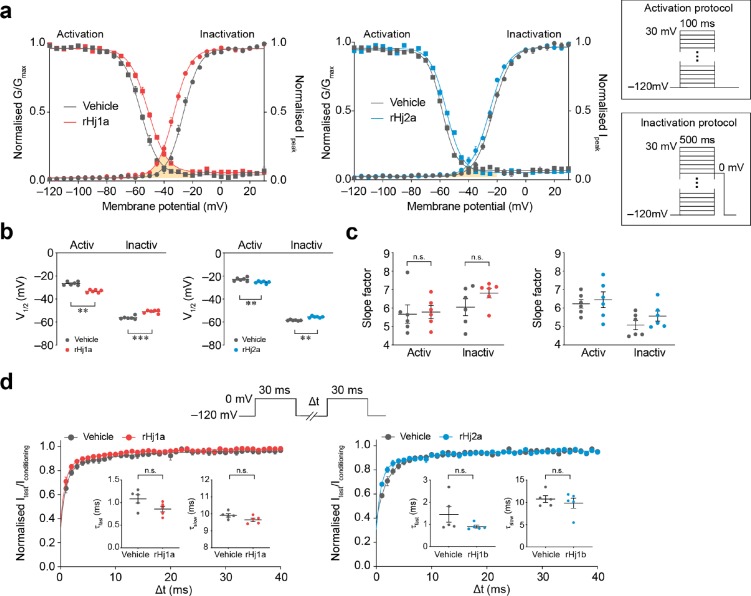
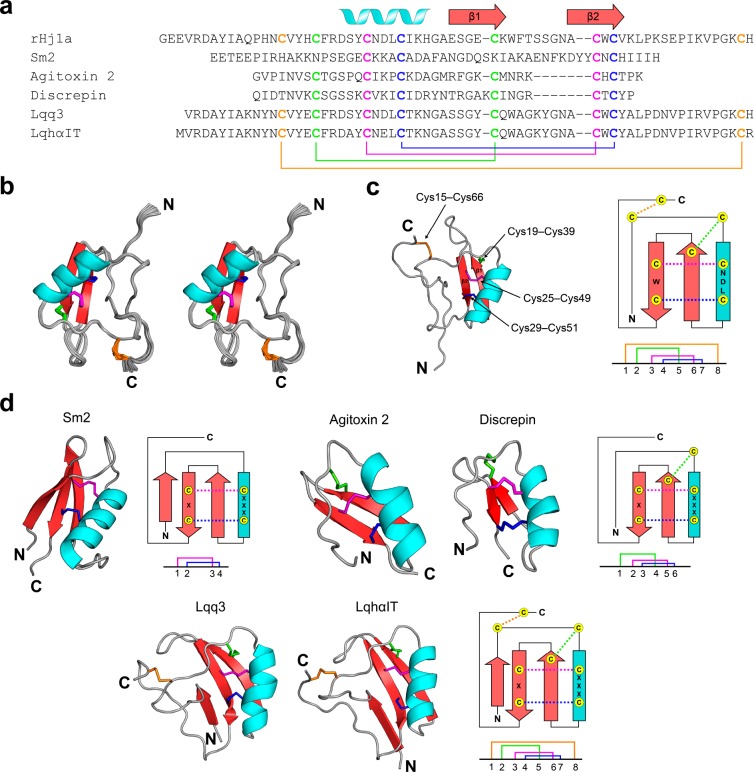
Voltage-gated sodium (NaV) channels play a fundamental role in normal neurological function, especially via the initiation and propagation of action potentials. The NaV1.1 subtype is found in inhibitory interneurons of the brain and it is essential for maintaining a balance between excitation and inhibition in neuronal networks. Heterozygous loss-of-function mutations of SCN1A, the gene encoding NaV1.1, underlie Dravet syndrome (DS), a severe pediatric epilepsy. We recently demonstrated that selective inhibition of NaV1.1 inactivation prevents seizures and premature death in a mouse model of DS. Thus, selective modulators of NaV1.1 might be useful therapeutics for treatment of DS as they target the underlying molecular deficit. Numerous scorpion-venom peptides have been shown to modulate the activity of NaV channels, but little is known about their activity at NaV1.1. Here we report the isolation, sequence, three-dimensional structure, recombinant production, and functional characterization of two peptidic modulators of NaV1.1 from venom of the buthid scorpion Hottentotta jayakari. These peptides, Hj1a and Hj2a, are potent agonists of NaV1.1 (EC50 of 17 and 32 nM, respectively), and they present dual α/β activity by modifying both the activation and inactivation properties of the channel. NMR studies of rHj1a indicate that it adopts a cystine-stabilized αβ fold similar to known scorpion toxins. Although Hj1a and Hj2a have only limited selectivity for NaV1.1, their unusual dual mode of action provides an alternative approach to the development of selective NaV1.1 modulators for the treatment of DS.


Conflict of interest statement

The authors declare no competing financial interest.

Figures

Figure 1
Figure 1
Identification of scorpion-venom peptides that agonize human NaV1.1. (a) Hottentotta jayakari, a buthid scorpion native to Iran, Oman, Saudi Arabia, United Arab Emirates, and Yemen (image courtesy of M. Hogan at the Florida State University). (b) Representative current traces for HEK293 cells expressing NaV1.1 in the presence of vehicle (gray) or H. jayakari venom (200 ng/μL; orange). Inward sodium currents were evoked using the voltage protocol shown above the traces. (c) Four groups of pooled fractions were screened against NaV1.1 using automated whole-cell patch clamp electrophysiology, and only group II (shaded orange) contained NaV1.1-active fractions. (d) Chromatograms resulting from subsequent purification of active peptides using a VisionHT HILIC column (left panel), followed by a diphenyl column (middle panel), and a Promix MP column (right panel). Asterisks denote active fractions. Native Hj1a (shaded red) and Hj2a (shaded blue) were purified to homogeneity. The acetonitrile gradient is indicated by a dotted line. (e) MS analysis revealed that these venom peptides have monoisotopic masses of 7476.30 Da (Hj1a, top panel) and 7112.20 Da (Hj2a, bottom panel). (f) Each of the isolated peptides recapitulated the NaV1.1-modulating activity of crude H. jayakari venom.
Figure 2
Figure 2
Sequence alignment of Hj1a and Hj2a with closely related α-scorpion toxins from Buthus martensii Karsch (BmKM1 and BmKm2), Buthus occitanus mardochei (Bom4), Leiurus quinquestriatus hebraeus (Lqh4 and LqhαIT), Leiurus quinquestriatus quinquestriatus (Lqq3 and Lqq4), Odonthobuthus doriae (OD1), and Mesobuthus eupeus (MeuNaTx-4 and MeuNaTx-5). Cysteine residues are shadowed in yellow and asterisks denote C-terminal amidation.
Figure 3
Figure 3
Overview of LC–MS/MS analysis of Hj1a. (a) Mass spectrum of reduced, alkylated, and undigested Hj1a. The Cys residues were alkylated with iodoethanol (indicated by C*). (b) MS1 spectrum of reduced and alkylated trypsin-digested Hj1a over the m/z range 300–1500. (c) Comparison of observed and theoretical monoisotopic masses for the ions observed, their corresponding residue positions, and fragment sequences. (d) MS2 spectrum of the doubly charged peptide ion at m/z 788.9 supports the presence of the sequence WFTSSGNACWCVK in Hj1a.
Figure 4
Figure 4
Overview of LC–MS/MS analysis of Hj2a. (a) Mass spectrum of reduced, alkylated, and undigested Hj2a. The Cys residues were alkylated with iodoethanol (indicated by C*). (b) MS1 spectrum of reduced and alkylated trypsin-digested Hj2a over the m/z range 400–1600. (c) Comparison of observed and theoretical monoisotopic masses for the ions observed, their corresponding residue positions and fragment sequences. (d) MS2 spectrum of doubly charged peptide ion at m/z 632.2 supports the presence of the sequence NSYCNNECTK in Hj2a.
Figure 5
Figure 5
Effect of rHj1a on human NaV1.1–1.7 expressed in HEK293 cells. (a) Representative currents for NaV1.1–1.7 in the presence of vehicle (gray) or 1 μM rHj1a (red). The shaded orange region indicates a sustained current at the end of a depolarizing pulse. (b) Relative size of NaV1.1–1.7 currents at 5 ms after the peak current following addition of 1 μM rHj1a (n = 5–8). (c) The sustained NaV1.4 and NaV1.5 currents after treatment with 1 μM rHj1a (22.1 ± 2.0% and 27.2 ± 2.5%, respectively) were 2.5-fold higher than NaV1.1 (8.8 ± 1.4%) (P < 0.0005, both). (d) Fitting the Hill equation to concentration–response curves for the sustained currents yielded EC50 values of 17.0 ± 1.9 nM (n = 6) for NaV1.1, 7.5 ± 1.2 nM (n = 8) for NaV1.4, 9.2 ± 0.8 nM (n = 6) for NaV1.5, and 37.3 ± 5.9 nM (n = 5) for NaV1.6. Sustained current (30 ms from peak current) was normalized to peak current to quantify the magnitude of the effect. Data are mean ± s.e.m. *P < 0.05, **P < 0.005, ***P < 0.0005, using one-way ANOVA followed by post hoc analysis using Tukey’s multiple comparisons test.
Figure 6
Figure 6
Effect of rHj2a on human NaV1.1–1.7 expressed in HEK293 cells. (a) Representative currents for NaV1.1–1.7 in the presence of vehicle (gray) or 1 μM rHj2a (blue). (b) Relative size of NaV1.1–1.7 currents at 5 ms after the peak current following addition of 1 μM rHj2a (n = 4–8). (c) The sustained NaV1.4 and NaV1.6 currents after treatment with 1 μM rHj1a (14.0 ± 1.9% and 14.0 ± 1.0%, respectively) were 5.5-fold higher than for NaV1.1 (2.5 ± 0.4%) (P < 0.0005, both). (d) Fitting the Hill equation to concentration–response curves for the sustained currents induced by rHj2a yielded EC50 values of 52.8 ± 2.5 nM (n = 5) for NaV1.1, 32.0 ± 7.5 nM (n = 6) for NaV1.4, 116.7 ± 23.5 nM (n = 8) for NaV1.5, 46.3 ± 6.2 nM (n = 5) for NaV1.6, and 147.4 ± 20.6 nM (n = 4) for NaV1.7. Sustained current (30 ms from peak current) was normalized to peak current to quantify the magnitude of the effect. Data are mean ± s.e.m. ***P < 0.0005, using one-way ANOVA followed by post hoc analysis using Tukey’s multiple comparisons test.
Figure 7
Figure 7
Effects of rHj1a and rHj2a on the biophysical properties of human NaV1.1. (a) Voltage dependence of normalized peak conductance (circles) and steady-state inactivation (squares) in the presence of vehicle (gray; n = 6), 1 μM rHj1a (red; n = 6) and 1 μM rHj2a (blue; n = 6). The shaded orange area represents the window current in the presence of peptide. The activation and inactivation protocols are shown in the inset. (b) Both peptides (1 μM) caused a significant hyperpolarizing shift in the voltage dependence of channel activation as well as a depolarizing shift in the voltage dependence of steady-state inactivation. (c) Neither peptide induced a change in the activation and inactivation slope factors. (d) Ratio of peak amplitude evoked by the test pulse to that evoked by the conditioning pulse, versus duration of interpulse interval (n = 5, both). The voltage protocol is shown in the upper panel. Curves represent a double-exponential fit, generating fast and slow recovery time constants (inset). Neither peptide (at 1 μM) induced a statistically significant change in either the fast or slow recovery time constants. Data are mean ± s.e.m. **P < 0.005, ***P < 0.0005; n.s., not significant, paired two-tailed Student’s t-test.
Figure 8
Figure 8
NMR solution structure of rHj1a. (a) Alignment of Hj1a with closely related CSαβ venom peptides from the centipede Scolopendra morsitans (Sm2) and the scorpions L. quinquestriatus hebraeus (agitoxin 2 and LqhαIT), Tityus discrepans (discrepin), and L. quinquestriatus quinquestriatus (Lqq3). Cysteine residues characteristic of CSαβ peptides are shown in bold. The secondary structural elements of rHj1a are shown above the sequence alignment. Disulfide-bond connectivities are colored coded and illustrated below the sequences. (b) Stereoview of the ensemble of 20 rHj1a structures (PDB code 6OHX) overlaid for best fit over the backbone atoms of residues 3–66. The N- and C-termini are labeled. (c) Schematic of the rHj1a structure determined in this study with the corresponding secondary structure topology on the right. The disulfide-bond connectivities are colored according to that in part a. (d) Structural variations of the CSαβ motif. Ribbon representations of centipede toxin Sm2 (PDB 6BL9), two short-chain scorpion toxins agitoxin 2 (PDB 1AGT) and discrepin (PDB 2AXK), and two long-chain scorpion toxins Lqq3 (PDB 1LQQ) and LqhαIT (PDB 1LQH), with the corresponding secondary structure topology and disulfide connectivities shown at right.

Similar articles

Cited by

References

    1. Catterall W. A. (2012) Voltage-gated sodium channels at 60: structure, function and pathophysiology. J. Physiol. 590, 2577–2589. 10.1113/jphysiol.2011.224204. - DOI - PMC - PubMed
    1. Catterall W. A. (2010) Ion channel voltage sensors: structure, function, and pathophysiology. Neuron 67, 915–928. 10.1016/j.neuron.2010.08.021. - DOI - PMC - PubMed
    1. Rubinstein M.; Westenbroek R. E.; Yu F. H.; Jones C. J.; Scheuer T.; Catterall W. A. (2015) Genetic background modulates impaired excitability of inhibitory neurons in a mouse model of Dravet syndrome. Neurobiol. Dis. 73, 106–117. 10.1016/j.nbd.2014.09.017. - DOI - PMC - PubMed
    1. Tai C.; Abe Y.; Westenbroek R. E.; Scheuer T.; Catterall W. A. (2014) Impaired excitability of somatostatin- and parvalbumin-expressing cortical interneurons in a mouse model of Dravet syndrome. Proc. Natl. Acad. Sci. U. S. A. 111, e313910.1073/pnas.1411131111. - DOI - PMC - PubMed
    1. Verret L.; Mann E. O.; Hang G. B.; Barth A. M.; Cobos I.; Ho K.; Devidze N.; Masliah E.; Kreitzer A. C.; Mody I.; Mucke L.; Palop J. J. (2012) Inhibitory interneuron deficit links altered network activity and cognitive dysfunction in Alzheimer model. Cell 149, 708–721. 10.1016/j.cell.2012.02.046. - DOI - PMC - PubMed

LinkOut - more resources