. 2020 Jun 27;29(10):1635-1647.
doi: 10.1093/hmg/ddaa059.

Ngly1 -/- rats develop neurodegenerative phenotypes and pathological abnormalities in their peripheral and central nervous systems

Affiliations

Ngly1 -/- rats develop neurodegenerative phenotypes and pathological abnormalities in their peripheral and central nervous systems

Makoto Asahina et al. Hum Mol Genet. .

Abstract

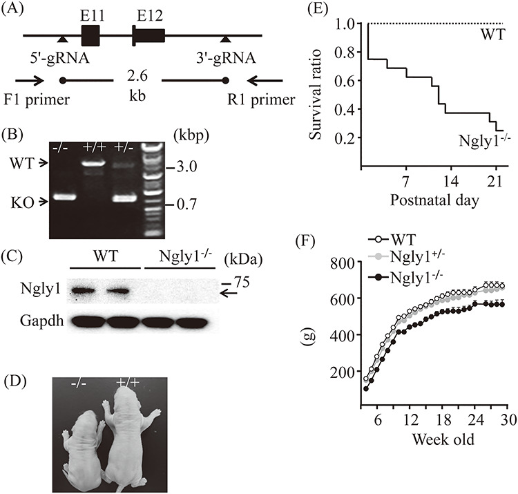
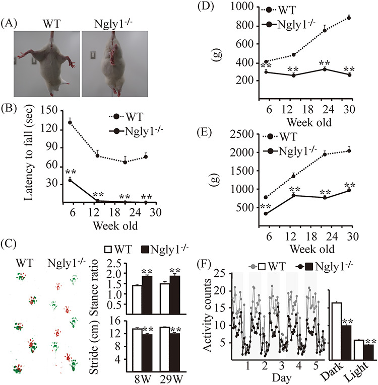
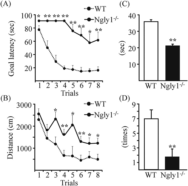
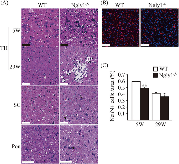
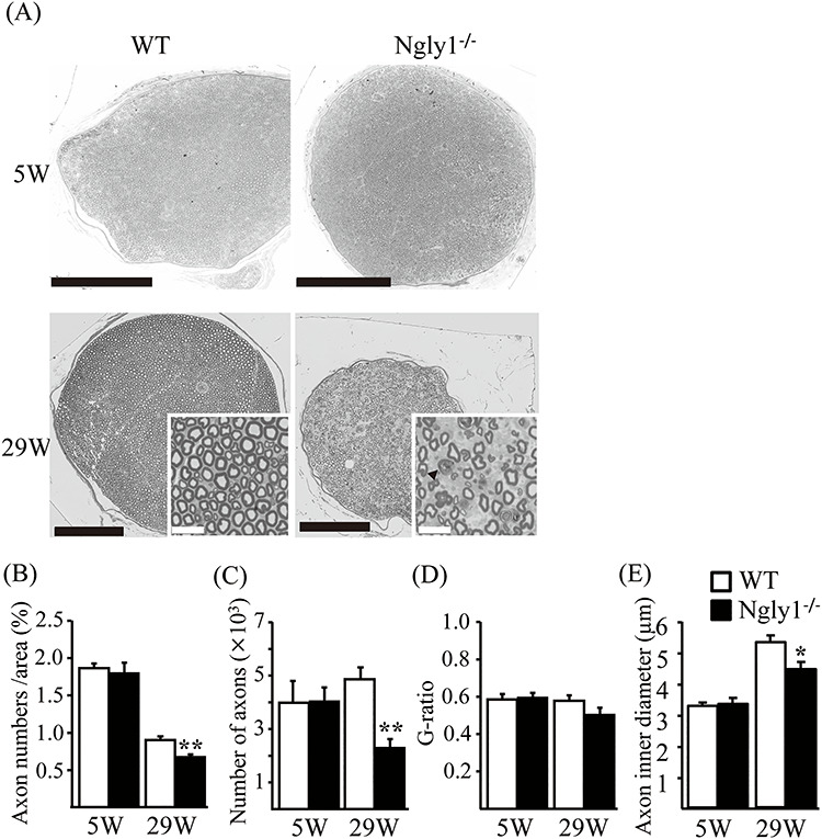
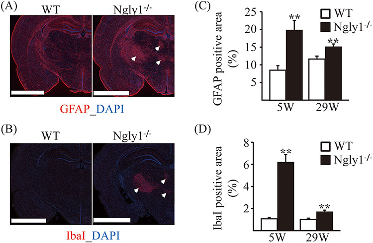
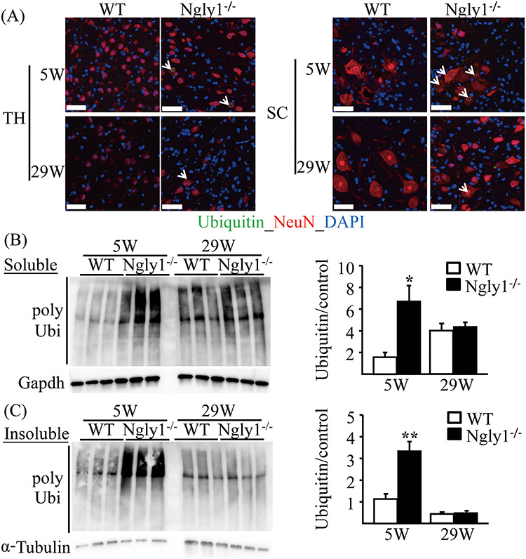
N-glycanase 1 (NGLY1) deficiency, an autosomal recessive disease caused by mutations in the NGLY1 gene, is characterized by developmental delay, hypolacrima or alacrima, seizure, intellectual disability, movement disorders and other neurological phenotypes. Because of few animal models that recapitulate these clinical signatures, the mechanisms of the onset of the disease and its progression are poorly understood, and the development of therapies is hindered. In this study, we generated the systemic Ngly1-deficient rodent model, Ngly1-/- rats, which showed developmental delay, movement disorder, somatosensory impairment and scoliosis. These phenotypes in Ngly1-/- rats are consistent with symptoms in human patients. In accordance with the pivotal role played by NGLY1 in endoplasmic reticulum-associated degradation processes, cleaving N-glycans from misfolded glycoproteins in the cytosol before they can be degraded by the proteasome, loss of Ngly1 led to accumulation of cytoplasmic ubiquitinated proteins, a marker of misfolded proteins in the neurons of the central nervous system of Ngly1-/- rats. Histological analysis identified prominent pathological abnormalities, including necrotic lesions, mineralization, intra- and extracellular eosinophilic bodies, astrogliosis, microgliosis and significant loss of mature neurons in the thalamic lateral and the medial parts of the ventral posterior nucleus and ventral lateral nucleus of Ngly1-/- rats. Axonal degradation in the sciatic nerves was also observed, as in human subjects. Ngly1-/- rats, which mimic the symptoms of human patients, will be a useful animal model for preclinical testing of therapeutic options and understanding the detailed mechanisms of NGLY1 deficiency.


Figures

Figure 1
Figure 1
Generation, appearance and body weights of Ngly1−/− rats. (A) Schematic figure showing generation of Ngly1−/− rat using CRISPR/Cas9 genome editing technology. (B) Representative results from genotyping of Ngly1+/+, +/− and −/− rats. Deletion in the Ngly1 gene can be detected by PCR and electrophoresis. (C) Western blot analysis of NGLY1 expression in brains from Ngly1−/− and WT rats. (D) Representative photograph of Ngly1−/− rat and a littermate WT rat at postnatal day one. (E) Survival curve of WT and Ngly1−/− rats (WT, n = 10; KO, n = 20). Ngly1−/− rats have a significantly shorter lifespan than their littermates, WT rats, until weaning. (F) Body weight of Ngly1−/−, Ngly1+/− and WT rats (Ngly1−/−; n = 10–12, Ngly1+/−; n = 12, WT; n = 12). Rats were weighed weekly after weaning (starting from 4 weeks old).
Figure 2
Figure 2
Motor dysfunction in Ngly1−/− rats. (A) Abnormal hindlimb clasping of Ngly1−/− rats compared with WT rats when suspended by the tail. (B) Rotarod testing for motor coordination of Ngly1−/− and WT rats at several ages. The time until drop from the accelerating rod (4–40 rpm in 4 min) is shown. (C) Gait analysis: left, representative paw placement records of 29-week-old rats; right, stride lengths and stance ratios of rats. (D, E) Grip-strength tests for assessment of forelimb (D) or forelimb and hindlimb (E) muscle force. (F) Home cage activity recorded over 120 h (left). The graph represents the means of 5-day averages of each analysis period (right). Values represent means ± SEM. The number of rats examined is 10–12 each. Asterisks indicate **P < 0.01, *P < 0.05.
Figure 3
Figure 3
Ngly1 −/− rats showed impaired spatial learning in Morris water maze tests. (A, B) Performance in eight trials during the acquisition stage of Ngly1−/− and WT rats. (A) Goal latency to reach the hidden platform (second). (B) Path length during each trial (centimeters). (C, D) Probe tests in the next day of the final acquisition trial. (C) Time spent in the quadrant (seconds). (D) The number of quadrant entries (mean ± SEM). The number of rats examined is 10 each. Asterisks indicate **P < 0.01, *P < 0.05.
Figure 4
Figure 4
Neuronal degeneration in the central nervous systems of Ngly1−/− rats. (A) H&E-stained sections of the thalamus (TH), spinal cords (SC) and pons from Ngly1−/− and the WT rats at 5 and 29 weeks of age. Black arrows indicate intra- and extracellular eosinophilic inclusion bodies and white arrows indicate mineralization. Black scale bar 50 μm, white scale bar 100 μm. (B) Immunohistochemistry of thalamus from Ngly1−/− and the WT rats at 5 weeks of age, stained with an anti-NeuN, a mature neuron marker (red). Nuclei were stained with DAPI. Scale bar 100 μm. (C) The number of NeuN-positive cells in VPM/VPL, VM and VL regions of the thalamus in Ngly1−/− and the WT rats at 5 and 29 weeks of age. Values represent mean ± SEM (n = 6–10). Asterisks indicate *P < 0.05 and **P < 0.01.
Figure 5
Figure 5
Axonal degeneration of older Ngly1−/− rats. (A) Toluidine-blue-stained plastic semi-thin sections of sciatic nerve of Ngly1−/− rats and WT rats at 5 or 29 weeks of age. Arrows indicate the myelin ovoid. Black scale bar 250 μm, white scale bar 25 μm. (B–E) Quantification of total number of axons (B), the density of axons (C), g-ratio (D) and average axon inner diameter (E) per nerve. Values represent mean ± SEM (n = 5–10). Asterisk, **P < 0.01, *P < 0.05 (Student’s t-test).
Figure 6
Figure 6
Glial activation in thalamic VPL/VPM, VM and VL regions of Ngly1−/− rats. Immunohistochemistry for GFAP in thalamus of WT and Ngly1−/− rats at 5 and 29 weeks of age, showing an increase in area of reactive astrocytes. (A, B) Immunohistochemistry for GFAP (A) and IbaI (B) in the thalamus of WT and Ngly1−/− rats at 5 weeks of age. Scale bar 2.5 mm. Arrows indicate the regions with gliosis in Ngly1−/− rats. Nuclei were stained with DAPI. (C, D) Quantitative analyses show the area occupied by the GFAP (C) or IbaI (D) positive areas in thalamus of WT and Ngly1−/− rats. Values represent mean ± SEM (n = 6). Asterisks indicate **P < 0.01.
Figure 7
Figure 7
Accumulation of ubiquitinated proteins in the central nervous systems of Ngly1−/− rats. (A) Ubiquitin-positive inclusions in neurons of Ngly1−/− rats. Immunohistochemistry of thalamus (TH) and spinal cord (SC) sections from Ngly1−/− and the WT rats at 5 and 29 weeks of age, stained with an anti-ubiquitin antibody and NeuN, a mature neuron marker. Arrows indicate ubiquitin-positive neurons. Nuclei were stained with DAPI. Scale bar 50 μm. (B, C) Accumulation of Triton-X-100-soluble (B) and insoluble (C) polyubiquitinated proteins in spinal cords of Ngly1−/− and WT rats. The total protein extracts were separated from the spinal cords of the rats and were separated into Triton-X-100-soluble and Triton-X-100-insoluble fractions and analyzed by immunoblotting using anti-polyubiquitinated antibodies (top) and anti-GAPDH or α-tubulin antibodies (bottom; loading control). Semi-quantitative analyses by densitometry were carried out (B, C). Values represent mean ± SEM (n = 3). Asterisks indicate *P < 0.05 and **P < 0.01.

References

    1. Suzuki T., Park H., Hollingsworth N.M., Sternglanz R. and Lennarz W.J. (2000) PNG1, a yeast gene encoding a highly conserved peptide:N-glycanase. J. Cell Biol., 149, 1039–1052. - PMC - PubMed
    1. Suzuki T., Huang C. and Fujihira H. (2016) The cytoplasmic peptide: N-glycanase (NGLY1) - structure, expression and cellular functions. Gene, 577, 1–7. - PMC - PubMed
    1. Hirsch C., Blom D. and Ploegh H.L. (2003) A role for N-glycanase in the cytosolic turnover of glycoproteins. EMBO J., 22, 1036–1046. - PMC - PubMed
    1. Huang C., Harada Y., Hosomi A., Masahara-Negishi Y., Seino J., Fujihira H., Funakoshi Y., Suzuki T., Dohmae N. and Suzuki T. (2015) Endo-β-N-acetylglucosaminidase forms N-GlcNAc protein aggregates during ER-associated degradation in Ngly1-defective cells. Proc. Natl. Acad. Sci. U. S. A., 112, 1398–1403. - PMC - PubMed
    1. Suzuki T. (2015) The cytoplasmic peptide:N-glycanase (Ngly1)-basic science encounters a human genetic disorder. J. Biochem., 157, 23–24. - PubMed

MeSH terms

Substances