. 2020 Apr 30;181(3):574-589.e14.
doi: 10.1016/j.cell.2020.03.004. Epub 2020 Apr 6.

An Airway Protection Program Revealed by Sweeping Genetic Control of Vagal Afferents

Affiliations

An Airway Protection Program Revealed by Sweeping Genetic Control of Vagal Afferents

Sara L Prescott et al. Cell. .

Abstract

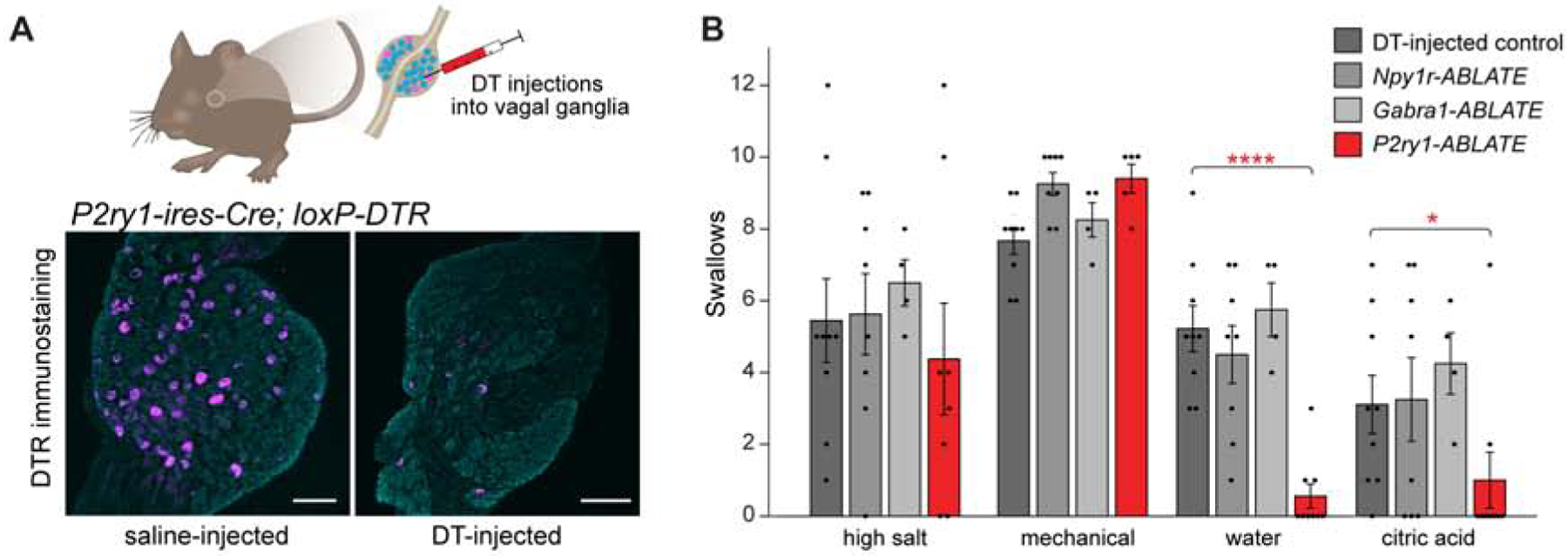
Sensory neurons initiate defensive reflexes that ensure airway integrity. Dysfunction of laryngeal neurons is life-threatening, causing pulmonary aspiration, dysphagia, and choking, yet relevant sensory pathways remain poorly understood. Here, we discover rare throat-innervating neurons (∼100 neurons/mouse) that guard the airways against assault. We used genetic tools that broadly cover a vagal/glossopharyngeal sensory neuron atlas to map, ablate, and control specific afferent populations. Optogenetic activation of vagal P2RY1 neurons evokes a coordinated airway defense program-apnea, vocal fold adduction, swallowing, and expiratory reflexes. Ablation of vagal P2RY1 neurons eliminates protective responses to laryngeal water and acid challenge. Anatomical mapping revealed numerous laryngeal terminal types, with P2RY1 neurons forming corpuscular endings that appose laryngeal taste buds. Epithelial cells are primary airway sentinels that communicate with second-order P2RY1 neurons through ATP. These findings provide mechanistic insights into airway defense and a general molecular/genetic roadmap for internal organ sensation by the vagus nerve.

Keywords: cough; interoception; larynx; nodose ganglion; optogenetics; peripheral nervous system; petrosal ganglion; purinergic receptors; single cell RNA sequencing; superior laryngeal nerve.


Conflict of interest statement

Declaration of Interests S.D.L. is a consultant for Kallyope.

Figures

Figure 1.
Figure 1.. Evoking swallows and expiratory reflexes with laryngeal stimulation and light
(A) Cartoon of upper airway innervation. (B) Schematic for perfusion of laryngeal stimuli. (C) Representative respiratory rhythms during (blue shading) delivery of isotonic saline (control), water, high salt (1500 mOsm) and citric acid (25 mM), with swallows (red triangles) and expiratory reflexes (blue triangles) noted. (D) Swallows (n=6–13, mean ± sem) were counted during laryngeal application of stimuli (see methods). (E) Swallows were counted before (control, gray) or after (SLN-cut, red) acute bilateral SLN transection (n=2–31, mean ± sem). (F) Cartoon (left) depicting optogenetics paradigm under urethane, and (right) changes in tracheal pressure (i: inspiration; e: expiration), digastric muscle electromyography (EMG), and pharyngeal pressure during a representative swallow and expiratory reflex. (G) Optogenetic stimulation (vagal trunk) in Vglut2-ires-Cre; loxP-Chr2 mice resulted in swallows (red triangles) and expiratory reflexes (blue triangles). Representative traces of tracheal pressure, digastric muscle EMG, and pharyngeal pressure over time are shown, yellow shading: light stimulation (20 Hz).
Figure 2.
Figure 2.. An atlas of sensory neuron subtypes from vagal and glossopharyngeal nerves
(A) Experimental pipeline for single-cell transcriptome analysis. (B) Uniform Manifold Approximation and Projection (UMAP) plots indicating cell subtype diversity across 25,117 NJP sensory neurons. Neuron clusters from jugular (J1-J10) and nodose/petrosal (NP1-NP27) ganglia are color coded. (C) Normalized expression levels (blue-gray scale) of cluster-defining signature genes (see Table S1 for genes 1–185) across all NJP sensory neurons analyzed. (D) UMAP plots from single-cell transcriptomes (left) and RNA in situ hybridization in cryosections of NJP ganglia (right) showing gene expression in sensory neuron subsets, scale bars: 100 μm. See also Figure S1 and Table S1.
Figure 3.
Figure 3.. A genetic toolkit for accessing small groups of NJP sensory neurons
(A) UMAP plots indicating expression of genes in NJP sensory neurons. (B) Native reporter fluorescence in cryosections of NJP ganglia from mice with Cre alleles and Cre-dependent reporter genes (L10-GFP, except tdTomato for Calb1-ires-Cre and Vglut-ires-Cre). tdTomato images were pseudocolored to match GFP images; far-red fluorescent Nissl counterstain (gray), scale bars: 100 μm. The image from Npy2r-ires-Cre mice was previously published (Chang et al., 2015). See also Figure S2.
Figure 4.
Figure 4.. P2RY1 neurons elicit a multi-faceted airway defense program
(A) Swallows and (B) expiratory reflexes were counted during vagus nerve optogenetics experiments in mice expressing ChR2 from Cre drivers indicated, mean ± sem. (C) UMAP plot indicating NP19 neurons, which are the only neurons that express P2ry1, but not Piezo2, Crhr2, Gpr65, or Gabra1. (D) Cartoon (top) depicting vocal fold dynamics, with images (bottom) of glottic area (green) during abduction and adduction, scale bar: 500 μm. (E) Representative graphs of glottic area over time during (yellow shading) vagus nerve optogenetics; triangles: swallows. (F) In vivo calcium imaging in NJP ganglia from 5 Piezo2-GCaMP* mice (left) and 3 P2ry1-GCaMP* mice (right). Rows indicate responses (ΔF/F, color scale) over time of individual neurons to lung stretch (15 seconds). Magenta and black bars represent tdTomato-positive and negative neurons. Not all unresponsive tdTomato-negative neurons are depicted; numbers at Y-axis base indicate total number of viable imaged neurons. See also Figure S3, S4.
Figure 5.
Figure 5.. P2RY1 neurons mediate responses to laryngeal water and acid challenge
(A) Cartoon (top) of DT-mediated ablation of Cre-expressing cells. DTR immunostaining of NJP ganglia from P2ry1-ires-Cre; loxP-DTR mice 3 weeks after intraganglionic saline (left) or DT (right) injection, far-red fluorescent Nissl counterstain (gray), scale bars: 100 μm. (B) Swallows over 20 second perfusion or 10 mechanical trials in mice indicated, n=4–9 mice, mean ± sem, *p<0.05, **p<0.005, ****p<0.00005. See also Figure S5.
Figure 6.
Figure 6.. P2RY1 neurons innervate laryngeal taste buds
(A) Cartoon depicting Cre-based anatomical mapping of vagal sensory neurons. (B) P2RY1 neuron terminals visualized in an open-book preparation of the larynx after injecting AAV-flex-AP into NJP ganglia of P2ry1-ires-Cre mice, scale bar: 500 μm. (C) Diagram depicting terminals of various neuron types in the larynx after AAV mapping. Red boxes i to vi show regions of larynx highlighted below. (D, E) Representative images of terminals formed by neuron subtypes captured by wholemount analysis of an open-book preparation (top) or in larynx cryosections (bottom) following tdTomato immunostaining, scale bars: 50 μm. (F) Taste buds visualized in larynx cryosections by KRT8 immunohistochemistry (green), scale bar: 100 μm. (G) Two-color immunohistochemistry for KRT8 (green) and tdTomato (magenta) in cryosections of larynx from P2ry1-ires-Cre mice injected with AAV-flex-tdTomato in NJP ganglia, scale bar: 50 μm. See also Figure S6.
Figure 7.
Figure 7.. Laryngeal reflexes involve epithelial cell-neuron communication
(A) Two-color immunohistochemistry for KRT8 (green) and eYFP (yellow) in cryosections of arytenoid epithelium from tamoxifen-treated Krt8-CreER; loxP-ChR2 mice (ChR2 encodes a ChR2-YFP fusion protein), scale bar: 100 μm. (B) Cartoon of epithelium optogenetics in Krt8-CreER; loxP-ChR2 mice, with illumination of oral cavity (1), larynx (2), trachea (3) or NJP ganglion. (C) Swallows per optogenetics trial (10 Hz) in tamoxifen-treated Krt8-CreER; loxP-ChR2 mice (n=3–9, mean ± sem), with light fiber positions indicated in (B). (D) Swallows evoked in wild type (control, n=13–31) and P2×2/P2×3−/− mice (n=6) by test stimuli, mean ± sem, **p<0.005, ***p<5×10⁻¹⁰. (E) Swallows evoked in wild type (control) and P2×2/P2×3−/− mice by laryngeal perfusion (20 seconds) of NaCl solutions of various concentration in water, n=8–31 control mice, 3–6 P2×2/P2×3−/− mice, mean ± sem, **p<0.005, ***p<5×10⁻¹⁰. See also Figure S7.

Comment in

References

    1. Abdulqawi R, Dockry R, Holt K, Layton G, McCarthy BG, Ford AP, and Smith JA (2015). P2×3 receptor antagonist (AF-219) in refractory chronic cough: a randomised, double-blind, placebo-controlled phase 2 study. Lancet 385, 1198–1205. - PubMed
    1. Anderson JW, Sant’Ambrogio FB, Mathew OP, and Sant’Ambrogio G (1990). Water-responsive laryngeal receptors in the dog are not specialized endings. Respir Physiol 79, 33–43. - PubMed
    1. Bai L, Mesgarzadeh S, Ramesh KS, Huey EL, Liu Y, Gray LA, Aitken TJ, Chen Y, Beutler LR, Ahn JS, et al. (2019). Genetic Identification of Vagal Sensory Neurons That Control Feeding. Cell 179, 1129–1143 e1123. - PMC - PubMed
    1. Baral P, Umans BD, Li L, Kirschbaum T, Wei Y, Zhou Y, Yipp BG, Liberles SD, and Chiu IM (2018). Nociceptor sensory neurons suppress neutrophil and gamma delta T cell responses in bacterial lung infections and lethal pneuomonia. Nature Medicine in press. - PMC - PubMed
    1. Bessac BF, and Jordt SE (2008). Breathtaking TRP channels: TRPA1 and TRPV1 in airway chemosensation and reflex control. Physiology (Bethesda) 23, 360–370. - PMC - PubMed

Publication types

Substances