. 2020 Jul 15;107(4):779-792.
doi: 10.1016/j.ijrobp.2020.03.026. Epub 2020 Apr 4.

Deletion of Mouse Setd4 Promotes the Recovery of Hematopoietic Failure

Affiliations

Deletion of Mouse Setd4 Promotes the Recovery of Hematopoietic Failure

Xing Feng et al. Int J Radiat Oncol Biol Phys. .

Abstract

Purpose: Acquired hematopoietic failure is commonly caused by therapeutic and accidental exposure of the bone marrow (BM) to toxic agents. Efficient recovery from BM failure is dictated not only by the intrinsic sensitivity and proliferation capacity of the hematopoietic stem and progenitor cells but also by the BM environment niche. Identification of genetic factors that improve recovery from hematopoietic failure is essential. Vertebrate SETD4 is a poorly characterized and putatively nonhistone methyltransferase. This study aims to identify the roles of SETD4 in BM recovery.

Methods and materials: An inducible SETD4 knockout mouse model (Setd4flox/flox;Rosa26-CreERT2+) was used. Adult sex-matched littermates were treated with tamoxifen to induce Setd4 deletion or oil as the control. Tamoxifen-treated Setd4wt/wt;Rosa26-CreERT2+ mice were included as another control. Those mice were irradiated to induce hematopoietic syndrome and analyzed to identify the roles and mechanisms of Setd4 in of BM recovery.

Results: Loss of Setd4 in adult mice improved the survival of whole-body irradiation-induced BM failure. This was associated with improved recoveries of long-term and short-term hematopoietic stem cells (HSCs) and early progenitor cells. BM transplantation analyses surprisingly showed that the improved recovery was not due to radiation resistance of the Setd4-deficient HSCs but that Setd4-deficient HSCs were actually more sensitive to radiation. However, the Setd4-deficient mice were better recipients for allogeneic HSC transplantation. Furthermore, there was enhanced splenic erythropoiesis in Setd4-deficient mice.

Conclusion: These findings not only revealed a previously unrecognized role of Setd4 as a unique modulator of hematopoiesis but also underscored the critical role of the BM niche in recovery from hematopoietic failure. Our study also implicated Setd4 as a potential target for therapeutic inhibition to improve the conditioning of the BM niche before allogeneic transplantation.


Conflict of interest statement

Conflict of Interest statement: The authors declare that there were no competing interests.

Figures

Figure 1.
Figure 1.. Sensitivity of Setd4 deficient mice to TBI.
(A&B): Tam or control treated sex-matched littermates of Setd4flox/flox;Rosa26-CreERT2+ or Setd4flox/wt;Rosa26-CreERT2+ were divided into three experiments groups: 1) Control: Oil-treated Setd4flox/flox;Rosa26-CreERT2+ or Setd4flox/wt;Rosa26-CreERT2+; 2) Setd4Δ/wt: Tam-treated Setd4flox/wt;Rosa26-CreERT2+; and 3) Setd4Δ/Δ: Tam-treated Setd4fox/flox;Rosa26-CreERT2+. Mice were treated with 8 Gy (A) or 9 Gy (B) of TBI, and monitored for survival. The P-values and number of mice in each group are shown. (C): Control mice treated with tamoxifen and Oil and then TBI as indicated. Oil: Oil-treated Setd4flox/flox;Rosa26CreERT2−/−, Setd4flox/wt;Rosa26CreERT2−/−, and Setd4wt/wt;Rosa26-CreERT2+ mice. For 8 Gy, 4, 3 and 2 mice of the above genotypes were included. For 9 Gy, 6, 2, and 2 mice were used. Tam: Tam-treated wt mice with the same genotypes above. For 8 Gy, 4, 4, and 4 mice with the same three genotypes were used. For 9 Gy, 6, 4, and 4 were included. The survivals of each group (panels A, B, and C) were obtained from multiple irradiation experiments of 1-5 pairs of mice per irradiation depending on the availability of matched littermates at the time of irradiation. Shown are the results of the combined number of mice. The total number of mice (n) and the number of males (M) and females (F) in each group are shown above the survival curves. (D): Repopulation of BM cells after TBI. At least 3 mice in each group were pathologically analyzed by hematoxylin-and-eosin (HE) staining. Shown are by representative images from the staining of cross sections of femur BM cavities from the indicated group of mice 14 days after 8 Gy of TBI. Hematopoietic cells clusters had dramatically repopulated the bone marrows of Tam-treated Setd4flox/flox;Rosa26CreERT2+ (Setd4Δ/Δ) mice, but not in Oil-treated Setd4flox/flox;Rosa26CreERT2+ (Setd4flox/flox) or the Tam-treated Setd4wt/wt;Rosa26-CreERT2+ (Tam-Setd4wt/wt). Upper panel, 40x; lower panel, 100x; scale bar = 100 μm. (E): The total number of BM mono-nuclear cell (BM-MNC) collected from one paired femur and tibia. Three independent experiments with an average of 3 mice per subset of time points were performed. The combined data are plotted.
Figure 2.
Figure 2.. Cell counts of hematopoietic stem cells (HSCs) and progenitor cells in the BM after 8 Gy of irradiation.
(A): A simplified illustration of the lineage hierarchy of the Lin cells. Based on Sca-1 and c-Kit expression, the Lin- cells were divided into three populations shown with different colors. In each population, the specific cell types can be identified by additional cell markers (see Supplemental Materials and Methods, and Fig. S4 for details). LSK: Sca-1+ and c-Kit+; LS K: Sca-1 and c-Kit+; LSlowKlow: Sca-1low c-Kitlow; LT-HSC: long-term HSC; ST-HSC: short-term HSC; MPP: multipotent progenitor; LMPP: lymphoid-primed MPPs; CMP: common myeloid progenitors; GMP: granulocyte-macrophage progenitors; MEP: megakaryocyte-erythroid progenitors. CLP: common lymphoid progenitors. (B-J): The numbers of total LSK, LT-HSC, ST-HSC, MPP, LMPP, CMP, MEP, GMP, and CLP cells at different the indicated times after 8 Gy of TBI. The BM-MNC (Fig. 1E) were then used to sort out the Lin- cells, and to determine the relative percentage of each of the cell types in the hematopoietic hierarchy as labeled above each panel. The designation of animal genotypes is shown in Panel B. For each tested mouse, a pair of femur and tibia were crushed to collect all BM cells at 4, 14, and 21 days after 8Gy of TBI. The mouse genotypes were: 1) Oil-treated Setd4flox/flox;Rosa26-CreERT2+ (Seid4flox/flox, blue filled cycle); 2) Tamoxifen-treated Setd4flox/flox;Rosa26-CreERT2+ (Setd4Δ/Δ red square); and 3) tamoxifen-treated Setd4wt/wt;Rosa26-CreERT2+ (Tam-Setd4wt/wt, filled black cycles). The mice in groups 1 and 2 were male littermates. In all panels, each dot represents the data from an independent mouse, and none-irradiated mice are shown as day 0. *: p<0.05, **: P<0.01. Shown are combined results from individual mice from 2-3 independent experiments with an average of 3 mice per time point in each experiment.
Figure 3.
Figure 3.. Enhanced splenic erythropoiesis in Setd4 deficient mice after 8 Gy of TBI.
The spleens were collected to analyze their cellular components. (A): Representative gross features of spleens, and spleen weight at 2 weeks post 8 Gy TBI from mice with the indicated genotypes. (B): Representative H&E staining of spleens at 2 weeks post 8 Gy TBI, scale bar: 100 μm. A denser population of cells were observed in the Setd4 deficient mice than the control mice. (C): A schematic representation of developmental sequence of erythroid cells from Stage 1 (SI) to Stage 5 (S5). HSC: hematopoietic stem cell; BFU-E: burst-forming unit-erythroid; CFU-E: colony-forming unit-erythroid; Pro-E: pro erythroblast; Chromo-E: chromatophilic erythroblasts; RBC: reticulocyte and erythrocyte. (D): Representative CD71 and TER119 staining profiling for spleen erythroid populations for the indicated mouse genotypes. The gating scheme to identify the different erythroid cell populations is shown on the right. (E): The numbers of total splenic erythroid cell populations (S2-S5) at 2 weeks post 8 Gy. Shown are combined quantification from two independent experiments. (F): Percentage distribution of B cells, T cells, granulocytes, and erythroid cells in the spleens at 2 weeks post 8 Gy for the indicated mouse genotypes.
Figure 4.
Figure 4.. Radiation sensitivity of wild type and Setd4 deficient BM chimeric mice.
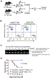
(A): Schema outlining the non-competitive BM reconstitution assay. B6.CD45.1 recipient mice (B6 mice expressing the congenic CD45.1 marker) were exposed to 2 split doses of 6.5 Gy (total of 13 Gy) separated by 4 hours to deplete their native BM. Then one million Lin– BM cells from sex-matched littermates of either Setd4flox/flox or Setd4Δ/Δ mice (with CD45.2 marker) were transplanted into recipient mice. Eight weeks after the transplantation, peripheral blood and ear clips were sampled to confirm the successful implantation of the donor BM. Sixteen weeks after the transplantation, the mice were exposed to 8 Gy of TBI to induce hematopoietic syndrome and to address whether there is any difference of sensitivity caused by the transplanted donor BM. (B): Representative flow cytometric profiles of peripheral blood cells from the B6.CD45.1 recipients reconstituted with CD45.2 Setd4flox/flox or CD45.2 Setd4Δ/Δ BM cells. The peripheral blood cells were sampled 8 weeks after BM transplantation, and then stained antibodies to differentiate the transplanted cells (CD45.2+) from the recipient cells (CD45.1+). The small percentage (6-7%) of the remaining CD45.1 host cells are likely radio-resistant that were not killed by the irradiation. (C): Conformation of the genotyping of the BM chimeric mice. Genomic DNA were extracted from ear snips (containing blood) of recipients mice that were reconstituted with Setd4Δ/Δ BM cells (lanes 1, 2, 3, 4, and 6) or with Setd4flox/flox BM cells (lanes 5, 7, 8). Multiplex PCR genotyping was performed to simultaneously identify the wild type, floxed exon 6, and ΔExon6 alleles using three mixed primers: A (5′-TCCTGGGCTCTGCCATCCATG), B (5′-CTGTTGCAATGGAAATGCCAG), and C (5′-CTAAAGCTCTGCCCTAAGGTC). Pairing between primers A and B can amplify a 234bp wt allele and a 318bp flox-Exon6 allele but cannot amplify the ΔExon6 allele because region of primer B is deleted in the ΔExon6 allele. Paring between primers A and C can amplify a 369bp ΔExon6 allele. (Details of the PCR genotype strategies can be found in the Supplement Information of a previous report (40). (D): Kaplan-Meier survival curves of BM chimeric mice after γ-radiation. Sixteen weeks after transplantation, the B6.CD45.1 recipients were treated with 8 Gy of TBI, and animal survival from the expected hemopoietic failure was monitored. Shown are the survival curves of the mice transplanted with Setd4Δ/Δ (n=13) or Setd4flox/flox (n=11). p = 0.0049.
Figure 5.
Figure 5.. Radiation sensitivity of engrafted HSCs from Setd4 deficient mice.
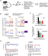
(A): Shema detailing the competitive repopulation assay. Lin– BM cells (>95% Lin–) from Setd4Δ/Δ mice (CD45.2) were mixed, at a ratio of 1:1, with WT competitor Lin–cells from B6.CD45.1/2 mice, and 1 × 10⁶ total cells were injected i.v. into lethally irradiated B6.CD45.1 recipients. The contribution of HSCs to reconstitute and subsequent refurbishment of the peripheral hematopoietic cells was monitored by flow cytometric analysis every 4 weeks. Twenty weeks after the transplantation, 6 of 11 reconstituted mice were exposed to sublethal irradiation (6.5 Gy) (IR: n=6; non-IR: n=5), and peripheral blood was monitored for 12 weeks, until final sacrifice for BM analysis at 18 weeks after the irradiation (or 48 weeks after the transplantation). (B): Peripheral blood from lethally irradiated transplanted mice were monitored for the relative contribution of Setd4Δ/Δ donor (CD45.2) and wild type competitor (CD45.1/2) cells every 4 weeks, starting 8 weeks post-transplantation. Shown are the ratio of CD45.2 cells over CD45.1/2 competitor cells in the total peripheral blood cells and the granulocytes. Data show mean ± SEM among the mice, and are representative of two identical experiments. See Table S4 for the number of animals in each group. The presented data were the combination of two independent experiments. (C): The relative contribution of irradiated Setd4Δ/Δ (donor) and WT (competitor) cells in all cells (left) or granulocytes (right) was plotted at 16, 24, 28 and 32 wk post transplantation. *, p < 0.05 by t-test. (D): The relative contribution of Setd4Δ/Δ and Setd4wt/wt BM HSCs and HPCs at 48 weeks after the transplantation. Left: irradiated mice (3 mice per group). Right: non-irradiated mice (2 mice per group). Mean and SEM are also shown. See Fig. S5 for representative flowcytometry profiles.
Figure 6.
Figure 6.. Survival of lethally irradiated Setd4 deficient and proficient mice after transplantation of wild type Lin- BM donor cells.
(A): Shema detailing HSC transplantation to rescue the lethally irradiated recipient mice. Serially diluted donor Lin- BM cells (10, 25, 40, 50, 60, 75, and 100x103) from B6.CD45.1 mice were transferred into Setd4Δ/Δ or Setd4flox/flox recipients that were irradiated with to 2 doses of 6.5 Gy separated by 4 hours (total of 13 Gy). Survival was monitored for 30 days. (B): Death rate of the transplanted recipient mice as a function of the numbers of transplanted Lin cells per mouse. For each experimental point, 6-14 recipients were used. The overall death rates were based on combined results from multiple independent transplantation experiments (see Table S5 for the specific number of mice used in each time point). The curves were fit by nonlinear regression. The calculated number of cells for a 63% of survival rate is 69.0x10³ for Setd4flox/flox mice, and 43.8x10³ for Setd4Δ/Δ mice. P=0.029 by two-way Anova test.

Similar articles

Cited by

References

    1. Pronk E, Raaijmakers MH. The mesenchymal niche in mds. Blood 2019;133:1031–1038. - PubMed
    1. Sieff CA. Introduction to acquired and inherited bone marrow failure. Hematology/Oncology Clinics 2018;32:569–580. - PubMed
    1. Bogliolo M, Surralles J. Fanconi anemia: A model disease for studies on human genetics and advanced therapeutics. Current opinion in genetics & development 2015;33:32–40. - PubMed
    1. Shimoni A, Nagler A. Radioimmunotherapy and stem-cell transplantation in the treatment of aggressive b-cell lymphoma. Leukemia & lymphoma 2007;48:2110–2120. - PubMed
    1. Calado RT. Immunologic aspects of hypoplastic myelodysplastic syndrome Seminars in oncology. Elsevier; 2011. pp. 667–672. - PMC - PubMed

Publication types