Review
. 2020 Jun:64:67-76.
doi: 10.1016/j.ceb.2020.02.015. Epub 2020 Apr 4.

Nuclear hubs built on RNAs and clustered organization of the genome

Affiliations
Review

Nuclear hubs built on RNAs and clustered organization of the genome

Kelly P Smith et al. Curr Opin Cell Biol. 2020 Jun.

Abstract

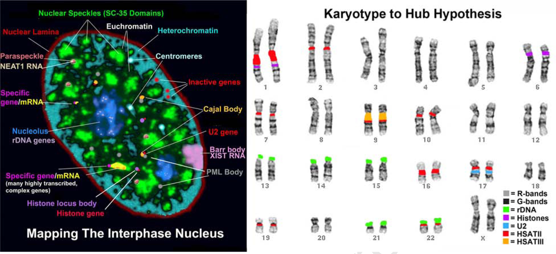
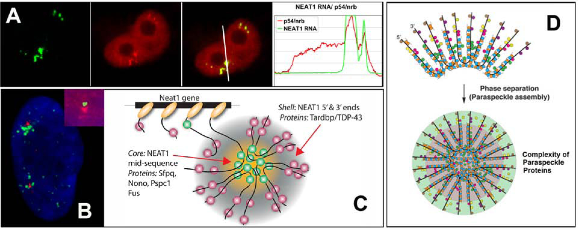
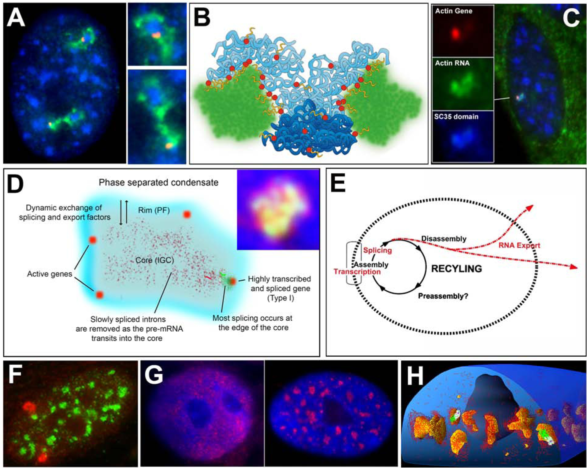
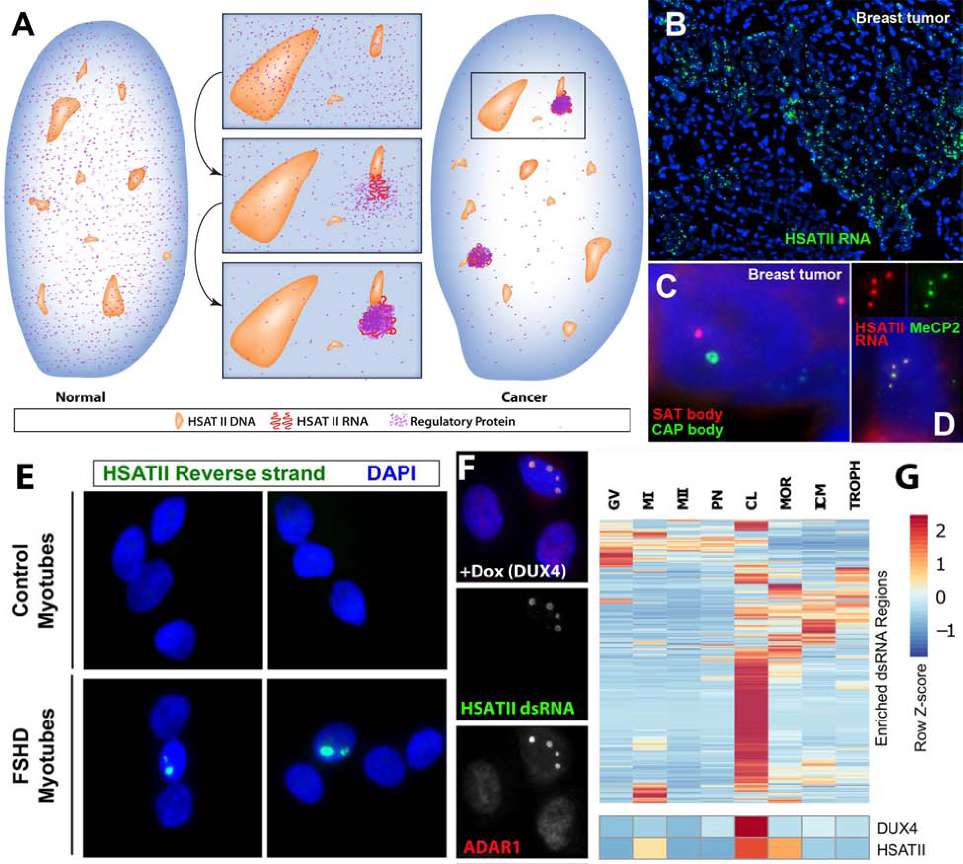
RNAs play diverse roles in formation and function of subnuclear compartments, most of which are associated with active genes. NEAT1 and NEAT2/MALAT1 exemplify long non-coding RNAs (lncRNAs) known to function in nuclear bodies; however, we suggest that RNA biogenesis itself may underpin much nuclear compartmentalization. Recent studies show that active genes cluster with nuclear speckles on a genome-wide scale, significantly advancing earlier cytological evidence that speckles (aka SC-35 domains) are hubs of concentrated pre-mRNA metabolism. We propose the 'karyotype to hub' hypothesis to explain this organization: clustering of genes in the human karyotype may have evolved to facilitate the formation of efficient nuclear hubs, driven in part by the propensity of ribonucleoproteins (RNPs) to form large-scale condensates. The special capacity of highly repetitive RNAs to impact architecture is highlighted by recent findings that human satellite II RNA sequesters factors into abnormal nuclear bodies in disease, potentially co-opting a normal developmental mechanism.

Keywords: Chromosome bands; Genome organization; Non-coding RNA; Nuclear structure; Speckles.


Conflict of interest statement

Conflict of interest statement Nothing declared.

Figures

Fig 1:
Fig 1:. Nuclear Compartmentalization Facilitated by Karyotype Organization
A) Mapping the Interphase nucleus: The nucleus contains many non-membrane bound sub-compartments defined by specific metabolic/regulatory factors, and the nuclear genome shows highly non-random organization relative to these. Active genes with similar RNA metabolic requirements often cluster with bodies or nuclear domains, which contain the many factors required for their biogenesis, creating efficient nuclear “hubs”. Most inactive heterochromatin positions close to the nuclear periphery (lamina) or the nucleolar periphery, whereas euchromatin largely positions more internally, where most factor-rich sub-structures form. This is a clear but flexible organization, such that not all active or inactive genes show the same organization. Nucleus composite shown is built on an image of DAPI DNA and SC-35 speckles in a human primary fibroblast nucleus. B) The Karyotype to Hub Hypothesis: We propose that human (and other) karyotypes evolved clustering of similar gene types to facilitate formation of efficient nuclear hubs, which in turn promote efficient gene expression. We point to several lines of evidence in the above karyotype which illustrate clustered genomic organization of genes with similar RNA metabolic requirements. These include the unique location of rDNA genes at the tips of all acrocentric chromosomes, the clusters of tandem U2 snRNA genes, and clustered organization of histone genes, all of which associate with specific nuclear bodies. We believe it self-evident that the exceptional organization of rDNA arrays at the tip of acrocentric chromosomes almost certainly evolved to enable these chromosomes to cluster and form the highly efficient nucleolar hub. Similarly, protein coding gene clustering on the chromosome arms likely facilitates gene clustering with nuclear speckles. The most dominant feature of human genome organization is the alternating light and dark bands as shown in this G-banded mitotic chromosome spread. For unknown reasons, the genome is organized in these alternating 2–10 Mb blocks (bands) of DNA with visibly distinct packaging (as seen with certain dyes), and substantial differences in gene and sequence content. Most genes localize in R-bands (Giemsa light bands in image), which are enriched in GC, SINE (Alu) repeats, and early replicating DNA. In contrast, dark G-bands are gene-poor, AT rich, LINE1 rich, and generally late replicating. Cytogenetic band patterns are essentially constant between people and cell-types, and the syntenic organization of genes shows substantial species conservation. As described in the text, other gene clusters (e.g. U2 snRNA or histone) also associate with Cajal or histone bodies which contain specific RNA metabolic factors. Also shown is the exceptionally high-copy ~26bp satellite repeat of HSATII (red), which includes a “mega-satellite” (~6MB) at Chr1q12, and smaller loci on several chromosomes. RNA from a large block of the 5bp HSATIII repeat (green, Chr9) is known to form heat-shock bodies to sequester factors in conditions of stress, analogous to recent evidence that HSATII satellites sequester factors in cancer and certain disease states.
Figure 2.
Figure 2.. NEAT1 is a Long Architectural RNA that Binds Many Proteins to Form Paraspeckles by Phase Separation.
A) Many proteins that are broadly distributed become enriched in paraspeckles (e.g. P54/nrb), but the NEAT1 RNA scaffold is exclusively found in these bodies [13]. B) Dense bodies of NEAT1 RNA (green) form as the RNA is transcribed from the locus (red, marked by the UHG locus at 11q13). Insert shows paraspeckles, enriched for PSP1 protein (red), emanate from the NEAT1 locus (green) in early G1 daughter cells before they distribute across the nucleus [13]. C) Model shows NEAT1 RNA building a phase separated paraspeckle coincident with transcription. The middle of the long transcript binds specific RBPs that form the inner core and are distinct from those that bind the 3’ and 5’ ends to form the outer shell. D) Model from Yamazaki et al 2018 [15] showing the complexity of the paraspeckle protein composition. NEAT1 has numerous repetitive sequences that bind many RBPs. This work demonstrates that long architectural RNAs are excellent scaffolds as they have the capacity to bind hundreds of proteins.
Figure 3.
Figure 3.. Genomic interactions, structure and function of nuclear speckles (SC-35 domains)
A) Fluorescence in situ hybridization (FISH) and immunocytochemical analysis shows that specific genes and gene rich R-bands associate with nuclear speckles. Both alleles of the Collagen 1A1 gene (red) associate with a speckle (blue) in essentially all human fibroblasts (where COL1A1 is ~4% of the total mRNA), while the closely linked R-band 17q22–24 (green) intimately associates with speckles. B) Model of speckle interactions with R and G-bands. (© 2003 Shopland et al. Originally published in the Journal of Cell Biology. https://doi.org/10.1083/jcb.200303131). Many, but not all, active genes (red) in the gene rich R-band (light blue) associate with nuclear speckles while gene poor G-bands (dark blue) exhibit less speckle association, and are more often at the nuclear periphery (as shown in fluorescence image in panel D). C) Many active genes, such as ß actin (ACTB) (red) associate with an SC-35 domain (blue), which corresponds to the speckle core (as shown in panel F). ACTB mRNA (green) distributes within the speckle core. D) While speckles were long believed to be merely storage sites, they are actually membraneless condensates that facilitate expression of associated genes through increased concentration and dynamic exchange of factors involved in mRNA metabolism. Many active genes (red) position in the rim (blue region), which corresponds to perichromatin fibril (PF) ultrastructures, where transcription occurs, but are adjacent to the speckle core (corresponding to interchromatin granule cluster (IGC) ultrastructures), which contains transcription, splicing and export factors. Most introns (green) of the COL1A1 gene were shown to be removed at the periphery at or near the gene, while a slow splicing intron (punctate red signals) distributed throughout the core (® 2000 Johnson et al. Originally published in the Journal of Cell Biology. https://doi.org/10.1083/jcb.150.3.417). Inset: Magnified image shows poly(A) RNA (red) defines the whole speckle and extends beyond the SC-35 defined core. (reproduced from: Hall et al. 2006). E) Model from Johnson et al, 2000 (Journal of Cell Biology. https://doi.org/10.1083/jcb.150.3.417) of a functional rationale for coupling the completion of mRNA maturation and release for mRNA export with the recycling/preassembly of RNA metabolic complexes in speckles. Since pre-mRNA processing requires interaction of many different factors, their concentration in condensates would facilitate recycling and reuse for expression of adjacent genes. F) Gene poor G-band at 3p14 (red) has minimal contact with speckles (green) and is closer to the nuclear periphery (reproduced from: Shopland et al. 2003, Journal of Cell Biology. https://doi.org/10.1083/jcb.200303131). G) Immunocytochemical detection of the speckle protein SC-35 (red) in human ES cells grown on mouse embryonic fibroblast feeders indicated that defined SC-35 rich speckles are largely absent in these samples of fully undifferentiated ES cells, although SC-35 staining was clearly detected through the nucleoplasm (left panel). SC-35 defined domains begin to coalesce during differentiation and appear well defined at day 7 (right panel) (Adapted from: Butler et al., 2009). H) Earlier 3D analysis in human fibroblasts showed that nuclear speckles organize in the same 3D plane within the nucleus, and associated genes and their pre-mRNAs (white and green) generally position adjacent in the X-Y plane, not the Z-axis. This organization is not simply due to the relatively flat nuclear morphology, as illustrated by the distinct positioning of the nucleolus. (From: Carter et al., 1993)
Figure 4.
Figure 4.. Long transcripts from high-copy tandem arrays have the marked capacity to sequester regulatory factors into nuclear bodies.
HSATII illustrates the exceptional capacity of high-copy satellite repeats to sequester factors at both the RNA and DNA level. Molecular cytology is required to demonstrate the distinct biology of different HSATII loci, which are divergent in the base 26 nt repeat sequence, are very poorly represented in reference genomes (only ~0.01% of 1q12 is in assembly GRCh38) and are very difficult to study using extraction-based methods. A) HSATII loci are not normally expressed in somatic cells, but many human tumors express HSATII RNA from a few smaller loci. The RNA accumulates into large satellite transcript (SAT) bodies that sequester regulatory factors and can thus affect gene regulation [66]. B) SAT bodies (green) are bright and prevalent, visible at low magnification in tumor sections. C) The large HSATII “mega satellite” at 1q12 does not express RNA in tumors studied, instead it accumulates repressive polycomb factors into CAP (Cancer Associated Polycomb) bodies that often co-exist with SAT-bodies in the same nucleus. D) HSATII RNA was found to sequester MeCP2 into SAT-bodies. (A-C from Hall et al 2017) E) In FSH dystrophy HSATII RNA again forms SAT-bodies in muscle (myotubes) linked to DUX4 expression. F) HSATII SAT-bodies in DUX4 expressing cells sequester ADAR1 and EIF4A3. G) DUX4 and HSATII are co-expressed normally in human cleavage stage embryos, indicating a likely normal developmental function for this long repetitive “junk” ncRNA. GV (germinal vesicle), MI (metaphase I), MII (metaphase II), PN (pronuclear stage), CL (cleavage stage), MOR (morula) and ICM (inner cell mass), TROPH (trophectoderm). (E-G from Shadle et al 2019).

Similar articles

Cited by

References

    1. Hall LL, Lawrence JB: RNA as a fundamental component of interphase chromosomes: could repeats prove key? Curr Opin Genet Dev 2016, 37:137–147. - PMC - PubMed
    1. Creamer KM, Lawrence JB: XIST RNA: a window into the broader role of RNA in nuclear chromosome architecture. Philos Trans R Soc Lond B Biol Sci 2017, 372. - PMC - PubMed
    1. Li X, Fu XD: Chromatin-associated RNAs as facilitators of functional genomic interactions. Nat Rev Genet 2019, 20:503–519. - PMC - PubMed
    1. Banani SF, Lee HO, Hyman AA, Rosen MK: Biomolecular condensates: organizers of cellular biochemistry. Nat Rev Mol Cell Biol 2017, 18:285–298. - PMC - PubMed
    1. Feric M, Vaidya N, Harmon TS, Mitrea DM, Zhu L, Richardson TM, Kriwacki RW, Pappu RV, Brangwynne CP: Coexisting Liquid Phases Underlie Nucleolar Subcompartments. Cell 2016, 165:1686–1697. - PMC - PubMed

Publication types