. 2020 Apr 3;21(7):2483.
doi: 10.3390/ijms21072483.

Female AhR Knockout Mice Develop a Minor Renal Insufficiency in an Adenine-Diet Model of Chronic Kidney Disease

Affiliations

Female AhR Knockout Mice Develop a Minor Renal Insufficiency in an Adenine-Diet Model of Chronic Kidney Disease

Camélia Makhloufi et al. Int J Mol Sci. .

Abstract

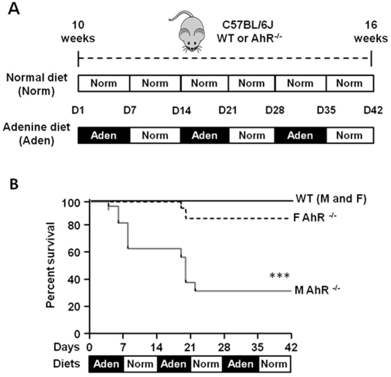
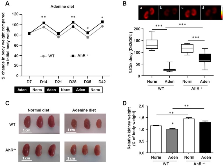
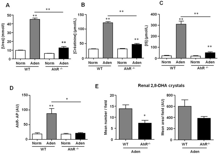
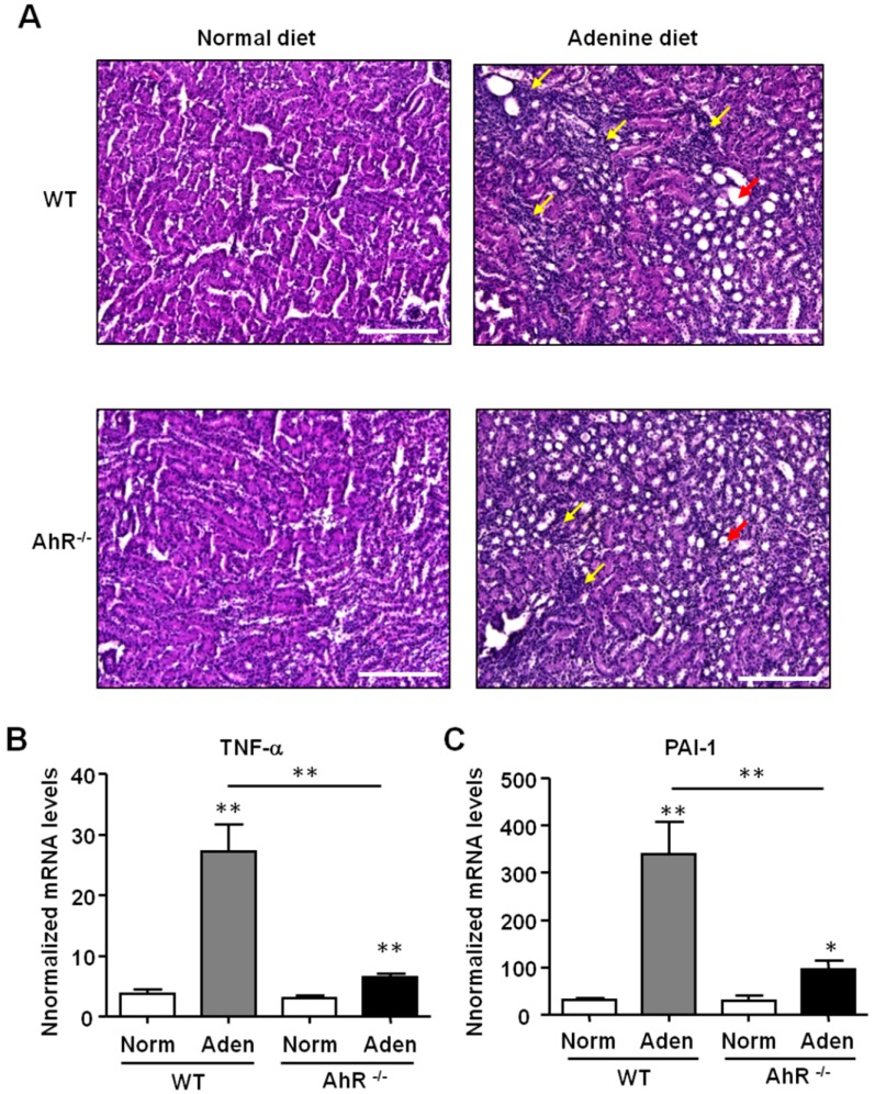
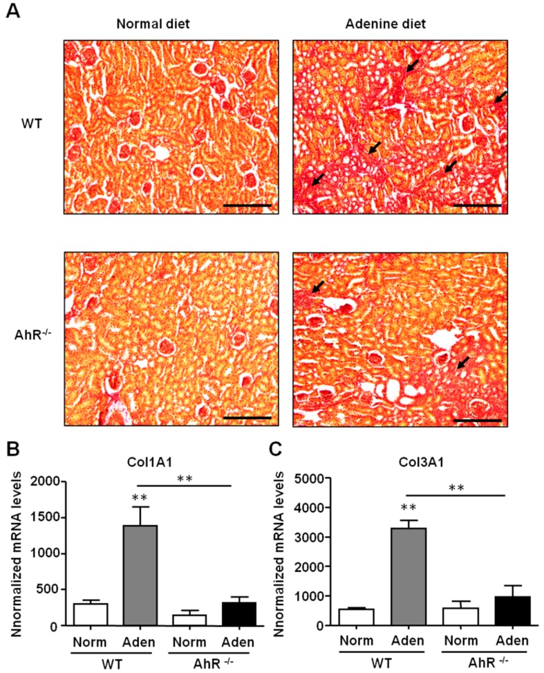
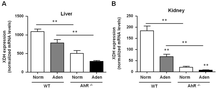
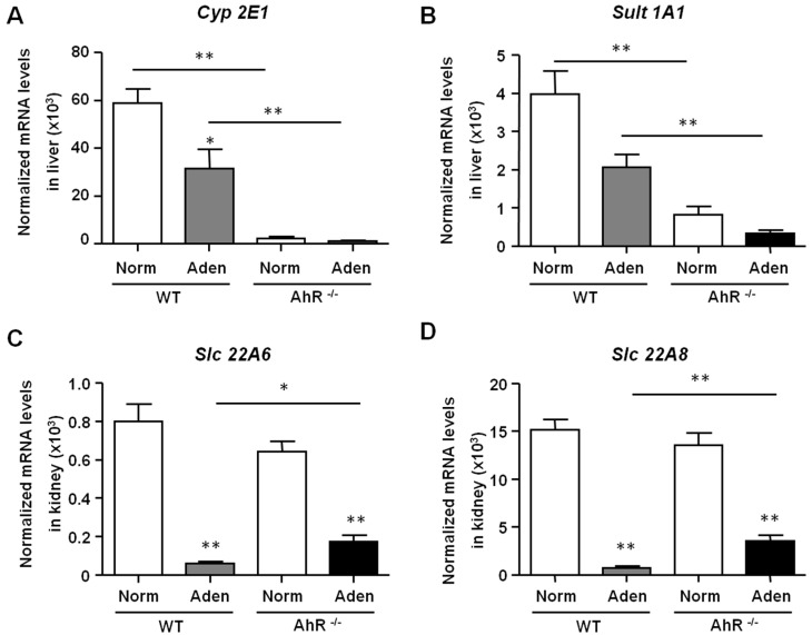
Cardiovascular complications observed in chronic kidney disease (CKD) are associated with aryl hydrocarbon receptor (AhR) activation by tryptophan-derived uremic toxins-mainly indoxyl sulfate (IS). AhR is a ligand-activated transcription factor originally characterized as a receptor of xenobiotics involved in detoxification. The aim of this study was to determine the role of AhR in a CKD mouse model based on an adenine diet. Wild-type (WT) and AhR-/- mice were fed by alternating an adenine-enriched diet and a regular diet for 6 weeks. Our results showed an increased mortality rate of AhR-/- males. AhR-/- females survived and developed a less severe renal insufficiency that WT mice, reflected by urea, creatinine, and IS measurement in serum. The protective effect was related to a decrease of pro-inflammatory and pro-fibrotic gene expression, an attenuation of tubular injury, and a decrease of 2,8-dihydroxyadenine crystal deposition in the kidneys of AhR-/- mice. These mice expressed low levels of xanthine dehydrogenase, which oxidizes adenine into 2,8-dihydroxyadenine, and low levels of the IS metabolism enzymes. In conclusion, the CKD model of adenine diet is not suitable for AhR knockout mice when studying the role of this transcription factor in cardiovascular complications, as observed in human CKD.

Keywords: adenine; aryl hydrocarbon receptor, indoxyl sulfate; chronic kidney disease; mouse model.


Conflict of interest statement

The authors declare no conflict of interest.

Figures

Figure 1
Figure 1
Effect on survival of wild-type (WT) and aryl hydrocarbon receptor (AhR)−/− mice fed by alternating an adenine-enriched diet and a regular diet for 6 weeks. (A) Timeline and experimental design for the normal diet (Norm) and adenine diet (Aden) supplied to C57BL/6J wild-type (WT) or AhR-knock out (KO) (AhR−/−) mice (10 weeks of age) for 42 days (D). (B) Kaplan–Meier survival curves of male (M) and female (F) WT and AhR−/− mice fed by alternating adenine-enriched (Aden) diet and regular diet (Norm) for 42 days (n = 13–16/group). *** p < 0.001 (log-rank test).
Figure 2
Figure 2
Body weight loss and renal impairment are less significant in AhR−/− mice following the adenine diet. (A) Percentage change in body weight compared to initial body weight of WT and AhR−/− mice fed for 42 days (D) alternatively with adenine-based chow (Aden) and normal chow (Norm). (B) Renal SPECT imaging with 99mTc-DMSA (dimercaptosuccinic acid) performed in WT and AhR−/− mice fed with a normal diet (a and c, respectively), or with an adenine (aden) diet (b and d, respectively). Results of 99mTc-DMSA uptake are expressed as box plots and represent the percentage ratio of percentages of injected dose (%ID) per kidney between day 42 and day 0 (D42/D 0%); n = 12 (6 mice/group). (C,D) Representative images and relative weight of left kidneys isolated from mice (WT and AhR−/−) fed with a normal diet (Norm) or an adenine-enriched diet (Aden). Data are expressed as mean ± SEM; n = 13–15/group (A) and n= 5–7/group (C). * p < 0.05; ** p < 0.01; *** p < 0.001.
Figure 3
Figure 3
AhR−/− mice are protected from renal insufficiency induced by an adenine diet. Urea (A), creatinine (B), and indoxyl sulfate (IS) (C) levels were determined in the serum of mice (WT and AhR−/−) from normal diet (Norm) and adenine diet (Aden) groups. (D) The AhR activating potential (AhR-AP) of serum was determined using the AhR-responsive CALUX (chemically activated luciferase expression) cell bioassay, as described in the material and methods. (E) The 2,8-DHA (dihydroxyadenine) crystals were quantified in sections of the right kidney, and results are expressed in mean number/field and mean area/field. Data are expressed as mean ± SEM, n = 13–15/group (A,B) and n = 5–7/group (CE). * p < 0.05; ** p < 0.01.
Figure 4
Figure 4
Adenine-induced renal insufficiency is associated with less pronounced inflammation in AhR−/− mice. (A) Representative hematoxylin and eosin (H&E) staining of kidney sections of mice (WT and AhR−/−) from the normal diet and adenine diet groups. The yellow arrows point areas of diffuse inflammation, and the red arrows indicate one dilated tubule. TNF-α (B) and PAI-1 (C) mRNA levels were determined in the kidneys of mice (WT and AhR−/−) obtained from the normal diet (Norm) and adenine diet (Aden) groups. Data are expressed as mean ± SEM, n = 5–7/group; * p < 0.05; ** p < 0.01. Scale bars = 200 μm.
Figure 5
Figure 5
Adenine-induced renal insufficiency is associated with less severe fibrosis in AhR−/− mice. (A) Representative picrosirius red staining of kidney sections of mice (WT and AhR−/−) from normal diet and adenine diet groups. The black arrows point to collagen deposition related to fibrosis. Col1A1 (B) and Col3A1 (C) mRNA levels were determined in the kidneys of mice (WT and AhR−/−) obtained from normal diet (Norm) and adenine diet (Aden) groups. Data are expressed as mean ± SEM, n = 5–7/group; ** p < 0.01. Scale bars = 200 µm.
Figure 6
Figure 6
AhR−/− mice had a lowest expression of XDH compared to WT mice. XDH mRNA levels were determined in the liver (A) and in kidneys (B) of mice (WT and AhR−/−) obtained from normal diet (Norm) and adenine diet (Aden) groups. Data are expressed as mean ± SEM, n = 5–7/group; * p < 0.05; ** p < 0.01.
Figure 7
Figure 7
Expression of hepatic enzymes of IS metabolism and IS renal transporters in WT and AhR−/− mice. Hepatic Cyp2E1 and Sult1A1 (A, B), as well as renal Slc22A6 and Slc22A8 (C,D) mRNA levels were determined in mice (WT and AhR−/−) obtained from normal diet (Norm) and adenine diet (Aden) groups. Data are expressed as mean ± SEM, n = 5–7/group; * p < 0.05; ** p < 0.01.

References

    1. Villain C., Metzger M., Combe C., Fouque D., Frimat L., Jacquelinet C., Laville M., Briancon S., Klein J., Schanstra J.P., et al. Prevalence of atheromatous and non-atheromatous cardiovascular disease by age in chronic kidney disease. Nephrol. Dial. Transplant. 2018 doi: 10.1093/ndt/gfy277. - DOI - PubMed
    1. Barreto F.C., Barreto D.V., Liabeuf S., Meert N., Glorieux G., Temmar M., Choukroun G., Vanholder R., Massy Z.A. Serum indoxyl sulfate is associated with vascular disease and mortality in chronic kidney disease patients. Clin. J. Am. Soc. Nephrol. 2009;4:1551–1558. doi: 10.2215/CJN.03980609. - DOI - PMC - PubMed
    1. Sallee M., Dou L., Cerini C., Poitevin S., Brunet P., Burtey S. The aryl hydrocarbon receptor-activating effect of uremic toxins from tryptophan metabolism: A new concept to understand cardiovascular complications of chronic kidney disease. Toxins (Basel) 2014;6:934–949. doi: 10.3390/toxins6030934. - DOI - PMC - PubMed
    1. Vanholder R., Schepers E., Pletinck A., Nagler E.V., Glorieux G. The uremic toxicity of indoxyl sulfate and p-cresyl sulfate: A systematic review. J. Am. Soc. Nephrol. 2014;25:1897–1907. doi: 10.1681/ASN.2013101062. - DOI - PMC - PubMed
    1. Addi T., Dou L., Burtey S. Tryptophan-Derived Uremic Toxins and Thrombosis in Chronic Kidney Disease. Toxins (Basel) 2018;10:412. doi: 10.3390/toxins10100412. - DOI - PMC - PubMed

LinkOut - more resources