. 2020 Apr 3;9(4):874.
doi: 10.3390/cells9040874.

Small Non-Coding RNA Profiling Identifies miR-181a-5p as a Mediator of Estrogen Receptor Beta-Induced Inhibition of Cholesterol Biosynthesis in Triple-Negative Breast Cancer

Affiliations

Small Non-Coding RNA Profiling Identifies miR-181a-5p as a Mediator of Estrogen Receptor Beta-Induced Inhibition of Cholesterol Biosynthesis in Triple-Negative Breast Cancer

Elena Alexandrova et al. Cells. .

Abstract

Triple-negative breast cancer (TNBC) is a highly heterogeneous disease, representing the most aggressive breast cancer (BC) subtype with limited treatment options due to a lack of estrogen receptor alpha (ERα), progesterone receptor (PR), and Erb-B2 receptor tyrosine kinase 2 (HER2/neu) expression. Estrogen receptor beta (ERβ) is present in a fraction of TNBC patients, where its expression correlates with improved patient outcomes, supported by the fact that it exerts oncosuppressive effects in TNBC cell models in vitro. ERβ is involved in microRNA-mediated regulation of gene expression in hormone-responsive BC cells and could mediate its actions through small noncoding RNAs (sncRNAs) in TNBCs also. To verify this possibility, smallRNA sequencing was performed on three ERβ-expressing cell lines from different TNBC molecular subtypes. Several sncRNAs resulted modulated by ERβ, with a subset being regulated in a tumor subtype-independent manner. Interestingly, sncRNA profiling of 12 ERβ+and 32 ERβ- primary TNBC biopsies identified 7 microRNAs, 1 PIWI-interacting RNA (piRNA), and 1 transfer RNA (tRNA) differentially expressed in ERβ+ compared to ERβ- tumors and cell lines. Among them, miR-181a-5p was found to be overexpressed in ERβ+ tumors and predicted target key components of the cholesterol biosynthesis pathway previously found to be inhibited by ERβ in TNBC cells.

Keywords: cholesterol biosynthesis; estrogen receptor beta; microRNA; small non-coding RNAs; triple-negative breast cancer.


Conflict of interest statement

The authors declare no conflict of interest.

Figures

Figure 1
Figure 1
Characterization of small noncoding RNA (sncRNA) expression profiles in triple-negative breast cancer (TNBC) cell lines. (A) Bar plot showing the number of expressed microRNAs (miRNAs), transfer RNA (tRNAs), small nucleolar RNAs (snoRNAs), small nuclear RNAs (snRNAs), PIWI-interacting RNA (piRNAs), and other small noncoding RNAs (sncRNAs) in the indicated cell lines. Only sncRNAs whose expression level exceeded three normalized reads are reported. (B) Venn diagram showing the number of common and specific miRNAs, highly expressed (expression level above the third quartile of the normalized read-count) in TNBC cell lines.
Figure 2
Figure 2
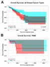
Estrogen receptor beta (ERβ) mRNA expression and survival of breast cancer patients from The Cancer Genome Atlas (TCGA) cohort [25]. Kaplan–Meier curves of overall survival for breast cancer (BC) (A) and TNBC (B) patients with respect to ERβ mRNA expression level (low expression: values below the first quartile, high expression: values above the third quartile). The highlighted area indicates confidence interval.
Figure 3
Figure 3
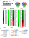
Effect of ERβ on sncRNA expression profiles and miRNA-regulated functions in TNBC. (A) Western blot analysis of ERβ expression in the HCC1806, MDA-MB-468, and Hs 578T cell lines upon induction of exogenous receptor expression by doxycycline (Doxy+) or in its absence (Doxy−). (B) Venn diagram of specific and commonly up-regulated (left panel) or down-regulated (right panel) sncRNAs in ERβ-expressing TNBC cells (|Fold-Change|(|FC|) ≥ 1.5, p < 0.05). Only sncRNAs characterized by the same behavior (up- or down-regulated) were included. (C) Heatmap showing ERβ-regulated miRNAs in the indicated cell lines (|FC| ≥ 1.5, p < 0.05) (D) Ingenuity Pathway Software (IPA) functional annotation analysis performed on ERβ-modulated genes predicted to be targets of differentially expressed miRNAs (|FC| ≥ 1.5, p < 0.05) in the corresponding cell lines. Commonly influenced functions in the three cell lines are indicated in orange. The vertical orange line indicates the p threshold (p < 0.05).
Figure 4
Figure 4
The ERβ-induced change in the sncRNA profile in TNBC tissues and its putative effect on molecular processes. (A) Histogram showing the number of miRNAs, tRNAs, snoRNAs, snRNAs, piRNAs, and other sncRNAs detected in ERβ− and ERβ+ tissue samples. Only sncRNAs whose median expression level exceeded a minimum threshold (three normalized reads) are reported. (B) Heatmaps showing miRNAs (left) and other sncRNAs (right) deregulated in ERβ+ TNBC tissues (|FC| ≥ 1.3, p < 0.05). RNAs differentially expressed in ERβ+ tumor biopsies and three or two cell lines are indicated in brown or orange, respectively. Graphic representation of statistically significant biological functions (C) and the top 20 statistically significant signaling pathways (D), identified by IPA performed on genes expressed in TNBC tissues and predicted to be targets of miRNAs commonly deregulated both in tissues and at least two of the studied cell lines.
Figure 5
Figure 5
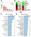
Putative effect of miR-181a-5p deregulation on molecular processes in TNBC tissues. Graphic representation of statistically significant biological functions (A) and the top 20 statistically significant signaling pathways (B), as identified by IPA performed on a group of genes expressed in TNBC tissues and predicted to be targets of miR-181a-5p. (C) Schematic representation of genes participating in cholesterol biosynthesis that were previously found down-regulated by ERβ in TNBC cells [15], with miR-181a-5p targets shown in red.

References

    1. Guo Y., Yu H., Wang J., Sheng Q., Zhao S., Zhao Y.Y., Lehmann B.D. The Landscape of Small Non-Coding RNAs in Triple-Negative Breast Cancer. Genes. 2018;9 doi: 10.3390/genes9010029. - DOI - PMC - PubMed
    1. Aysola K., Desai A., Welch C., Xu J., Qin Y., Reddy V., Matthews R., Owens C., Okoli J., Beech D.J., et al. Triple Negative Breast Cancer—An Overview. Hered. Genet. 2013;2013 doi: 10.4172/2161-1041.S2-001. - DOI - PMC - PubMed
    1. Foulkes W.D., Smith I.E., Reis-Filho J.S. Triple-negative breast cancer. N. Engl. J. Med. 2010;363:1938–1948. doi: 10.1056/NEJMra1001389. - DOI - PubMed
    1. Andreopoulou E., Schweber S.J., Sparano J.A., McDaid H.M. Therapies for triple negative breast cancer. Expert Opin. Pharmacother. 2015;16:983–998. doi: 10.1517/14656566.2015.1032246. - DOI - PMC - PubMed
    1. Boichuk S., Galembikova A., Sitenkov A., Khusnutdinov R., Dunaev P., Valeeva E., Usolova N. Establishment and characterization of a triple negative basal-like breast cancer cell line with multi-drug resistance. Oncol. Lett. 2017;14:5039–5045. doi: 10.3892/ol.2017.6795. - DOI - PMC - PubMed

Publication types

MeSH terms