. 2020 Apr 3;10(4):546.
doi: 10.3390/biom10040546.

Ranunculus bulumei Methanol Extract Exerts Anti-Inflammatory Activity by Targeting Src/Syk in NF-κB Signaling

Affiliations

Ranunculus bulumei Methanol Extract Exerts Anti-Inflammatory Activity by Targeting Src/Syk in NF-κB Signaling

Yo Han Hong et al. Biomolecules. .

Abstract

(1) Background: Ranunculus bulumei is a flowering plant that belongs to the Ranunculus species. Several Ranunculus species, such as R. aquatilis and R. muricatus, have traditionally been used to treat fever and rheumatism throughout Asia, suggesting that plants belonging to the Ranunculus species may have anti-inflammatory effects. To our knowledge, the pharmacological activity of R. bulumei has not been reported. Therefore, in this study, we aim to assess the anti-inflammatory activity of a methanol extract that was derived from R. bulumei (Rb-ME) in macrophage-mediated inflammatory responses and to identify the molecular mechanism that underlies any anti-inflammatory action. (2) Methods: The anti-inflammatory efficacy of Rb-ME was evaluated while using in vitro and in vivo experiments. The RAW264.7 cells and peritoneal macrophages were stimulated by lipopolysaccharide (LPS). In addition, LPS-induced peritonitis and HCl/EtOH-triggered gastritis models were produced. A nitric oxide (NO) assay, real-time PCR, luciferase reporter gene assay, western blot analysis, plasmid overexpression strategy, and in vitro kinase assay were used to determine the molecular mechanisms and target molecules of Rb-ME. The phytochemical active ingredients of Rb-ME were also identified by high performance liquid chromatograph (HPLC). (3) Results: Rb-ME reduced the production of NO and mRNA expression of iNOS, COX-2, IL-1β, and IL-6 without cytotoxicity. The protein secretion of TNF-α and IL-6 was also decreased by Rb-ME. HPLC analysis indicates that quercetin, luteolin, and kaempferol are the main active ingredients in the anti-inflammatory efficacy of Rb-ME. Rb-ME also blocked MyD88-induced NF-κB promoter activity and nuclear translocation of NF-κB subunits (p65 and p50). Moreover, Rb-ME reduced the phosphorylation of IκBα, Akt, p85, Src, and Syk, which are NF-κB upstream signaling molecules in LPS-activated RAW264.7 cells. According to the in vitro kinase assay, Rb-ME directly inhibits Syk kinase activity. The oral administration of Rb-ME alleviated inflammatory responses and the levels of p-IκBα in mice with LPS-induced peritonitis and HCl/EtOH-induced gastritis. (4) Conclusions Rb-ME has anti-inflammatory capacity by suppressing NF-κB signaling and it has been found to target Src and Syk in the NF-κB pathway. Based on this efficacy, Rb-ME could be developed as an anti-inflammatory herbal medicine.

Keywords: NF-κB signal pathway; Ranunculus bulumei; Src; Syk; anti-inflammatory activity.


Conflict of interest statement

The authors declare no conflict of interest.

Figures
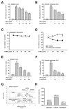
Figure 1
Figure 1
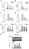
Inhibitory effect of R. bulumei (Rb-ME) on nitric oxide (NO) production in macrophages. (A and B) LPS-stimulated RAW264.7 cells (A) and peritoneal macrophages (B) were analyzed by NO assay in the presence or absence of Rb-ME. (C and D) Viability of Rb-ME-treated RAW264.7 cells (C), peritoneal macrophage, and HEK 293 cells (D) were measured by 3-(4-5-Dimethylthiazol-2-yl)-2-5-diphenyltetrazolium bromide (MTT) assay. (E and F) Levels of NO production in Pred-treated RAW264.7 cells (E) and L-NAME-treated peritoneal macrophages (F) in indicated doses were assessed by using a NO assay. (G and H) Active phytochemical constituents present in Rb-ME have been identified by HPLC. The profiles of Rb-ME components were analyzed by comparison with profiles obtained from standard samples of quercetin, luteolin, and kaempferol, which consisted of area and concentration. The data presented in (A), (B), (C), (D), (E), and (F) are shown as the means ± SD of two independent experiments and each experimental group was performed with ten (10) parallel wells to ensure the reliability of the results. ## p < 0.01 compared to untreated group, and * p < 0.05 and ** p < 0.01 compared to control group treated with LPS alone.
Figure 2
Figure 2
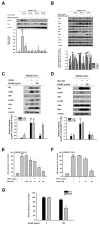
Effect of Rb-ME on the expression of inflammatory biomarkers at a transcriptional level. (A and B) RAW264.7 cells were pre-treated with Rb-ME (0–100 µg/mL) for 30 min. and then stimulated with LPS (1 µg/mL) for 6 h. The mRNA expression levels of proinflammatory cytokines, including iNOS, COX-2, TNF-α IL-1β and IL-6, were measured using semi-quantitative RT-PCR. (C and D) RAW264.7cells were pretreated with doses of Rb-ME (0-100 µg/mL) for 30 min. and then additionally treated with LPS (1 µg/mL) for 24 h, respectively. Protein levels of (C) TNF-α and (D) IL-6 released from the RAW264.7 cells were determined by ELISA kit. (E and F) HEK 293 cells were transfected with NF-κB-Luc and β-gal constructs with Flag-MyD88 or CFP-TRIF overexpressed (E). Flag-MyD88 or CFP-TRIF-transfected HEK 293 cells were overexpressed with AP-1-Luc and β-gal (F). Then, HEK 293 cells were additionally treated with Rb-ME (0–100 µg/mL) for 24 h. Luciferase activity was normalized using β-gal values after luminometer measurements. (G) Rb-ME (0 or 100 μg/mL) treated RAW264.7 cells were stimulated with LPS for the indicated times. Western blot analysis was performed with nuclear fractions to determine the nuclear translocation levels of NF-κB subunits (p65 and p50). Lamin A/C was used as a loading control for the nuclear fraction. The data presented at (A) and (B) are expressed as the means ± SD of three independent experiments. The data presented in (C), (D), (E) and (F) are expressed as the means ± SD of two independent experiments and each experimental group was performed with six (6) parallel wells to ensure the reliability of the results. The data presented in (G) is a representative of two independent experiments. # p < 0.05 and ## p < 0.01 compared to untreated group, and * p < 0.05 and ** p < 0.01 compared to control groups treated with LPS, MyD88 or TRIF alone.
Figure 3
Figure 3
Suppressive effect of Rb-ME on the NF-κB signal pathway. (A and B) Western blotting was performed using the whole lysates obtained from LPS-treated RAW264.7 cells for the indicated time in the presence or absence of Rb-ME (100 μg/mL). The levels of phosphorylated and total forms of IκBα (A), and Src, Syk, p85, Akt, IκBα, and β-actin (B) were determined by using specific antibodies for phospho- or total-proteins. (C and D) HEK 293 cells were transfected with HA-Src (C) or Myc-Syk (D), followed by treatment with Rb-ME (100 μg/mL) for 24 h. Src, Syk, HA, Myc, and β-actin, as well as phospho- or total-forms of p85 and IκBα were assessed by immunoblotting. (E and F) NO production was analyzed by a NO assay in LPS-stimulated RAW264.7 cells after treatment with PP2, a Src inhibitor (E), or Picea, a Syk inhibitor (F). (G) The effects of Rb-ME on Src and Syk activity were examined using an in vitro kinase assay with purified Src and Syk. The data presented in (A), (B), (C), and (D) are a representative of two independent experiments. Relative intensity is expressed as means ± SD of data measured and quantified using image J. The data presented in (E) and (F) are expressed as the means ± SD of two independent experiments and each experimental group was performed with ten (10) parallel wells to ensure the reliability of the results. # p < 0.05 and ## p < 0.01 compared to untreated group, and * p < 0.05 and ** p < 0.01 compared to control groups treated with LPS, Src, or Syk alone.
Figure 4
Figure 4
Anti-inflammatory effect of Rb-ME in peritonitis and gastritis models in vivo. (A and B) Rb-ME (50 and 100 mg/kg) was orally administered to C57BL/6 mice once a day for 5 days, and peritonitis was induced by intraperitoneal injection of LPS (10 μg/kg) for 1 day before mice were sacrificed. The effect of Rb-ME on NO production was analyzed in peritoneal exudates by using a NO assay (A). The number of leukocytes in peritoneal exudates was counted by using and a Neubauer chamber after staining with Turk solution (B). (C and D) Rb-ME (50 and 100 mg/kg) or ranitidine (40 mg/kg) was orally administered to ICR mice two times a day for two days. Gastritis was induced by HCl/EtOH injection 1 h before sacrifice. Gastric inflammatory lesions were taken with a digital camera and then quantitatively measured using Image j (C). Myeloperoxidase (MPO) activity was determined in total lysates obtained from the stomach tissue of gastritis mice treated with Rb-ME or ranitidine using an MPO activity colorimetric assay kit (D). (E and F) Immunoblotting assay was performed with whole lysates obtained from peritoneal exudates of peritonitis mice (D) or stomach tissue of gastritis mice (E). Phospho- or total-forms of IκBα and β-actin were detected using specific antibodies. The data presented in (A), (B), (C), and (D) are expressed as the mean ± SD of experiments that were performed with ten (A and B) and four (C and D) mice per group. The data presented in (E) and (F) are a representative of two independent experiments. Relative intensity is expressed as means ± SD of data measured and quantified using image J. # p < 0.05 and ## p < 0.01 as compared to untreated group, and * p < 0.05 and ** p < 0.01 compared to control groups treated with LPS or HCl/EtOH alone.
Figure 5
Figure 5
Schematic diagram of the anti-inflammatory effect of Rb-ME through inhibition of NF-κB signaling.

References

    1. Fujiwara N., Kobayashi K. Macrophages in inflammation. Curr. Drug Target Inflamm. Allergy. 2005;4:281–286. doi: 10.2174/1568010054022024. - DOI - PubMed
    1. Akira S., Uematsu S., Takeuchi O. Pathogen Recognition and Innate Immunity. Cell. 2006;124:783–801. doi: 10.1016/j.cell.2006.02.015. - DOI - PubMed
    1. Medzhitov R., Preston-Hurlburt P., Janeway C.A. A human homologue of the Drosophila Toll protein signals activation of adaptive immunity. Nature. 1997;388:394–397. doi: 10.1038/41131. - DOI - PubMed
    1. Correia J.D.S., Soldau K., Christen U., Tobias P.S., Ulevitch R.J. Lipopolysaccharide Is in Close Proximity to Each of the Proteins in Its Membrane Receptor Complex. J. Boil. Chem. 2001;276:21129–21135. doi: 10.1074/jbc.M009164200. - DOI - PubMed
    1. Ullah M.O., Sweet M.J., Mansell A., Kellie S., Kobe B. TRIF-dependent TLR signaling, its functions in host defense and inflammation, and its potential as a therapeutic target. J. Leukoc. Boil. 2016;100:27–45. doi: 10.1189/jlb.2RI1115-531R. - DOI - PubMed

Publication types