Review
. 2020 Apr 4;13(7):1688.
doi: 10.3390/ma13071688.

3D Printing of Hierarchical Scaffolds Based on Mesoporous Bioactive Glasses (MBGs)-Fundamentals and Applications

Affiliations
Review

3D Printing of Hierarchical Scaffolds Based on Mesoporous Bioactive Glasses (MBGs)-Fundamentals and Applications

Francesco Baino et al. Materials (Basel). .

Abstract

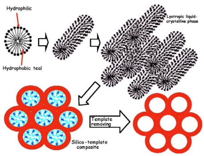
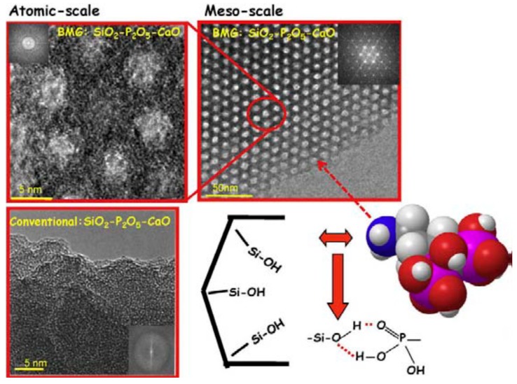
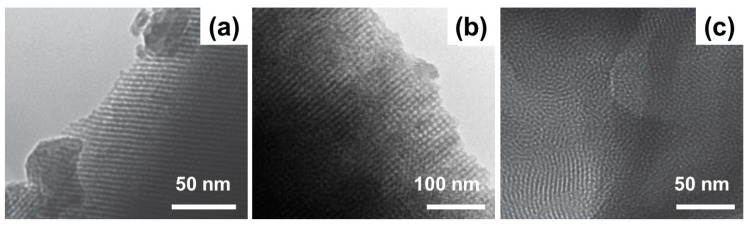
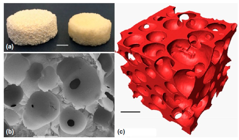
The advent of mesoporous bioactive glasses (MBGs) in applied bio-sciences led to the birth of a new class of nanostructured materials combining triple functionality, that is, bone-bonding capability, drug delivery and therapeutic ion release. However, the development of hierarchical three-dimensional (3D) scaffolds based on MBGs may be difficult due to some inherent drawbacks of MBGs (e.g., high brittleness) and technological challenges related to their fabrication in a multiscale porous form. For example, MBG-based scaffolds produced by conventional porogen-assisted methods exhibit a very low mechanical strength, making them unsuitable for clinical applications. The application of additive manufacturing techniques significantly improved the processing of these materials, making it easier preserving the textural and functional properties of MBGs and allowing stronger scaffolds to be produced. This review provides an overview of the major aspects relevant to 3D printing of MBGs, including technological issues and potential applications of final products in medicine.

Keywords: additive manufacturing; bioactivity; bioglass; biomaterials; drug delivery; hierarchical; ion release; mesoporous; porosity; scaffold; tissue regeneration.


Conflict of interest statement

The authors declare no conflict of interest.

Figures

Figure 1
Figure 1
Schematic illustration of the steps leading to a mesoporous silicate material starting from a micellar solution: micelles from structure directing agent (SDA) link the hydrolyzed silica precursors through the hydrophilic component and self-assembly to form an ordered mesophase. Pictures reproduced from Arcos and Vallet-Regi [16] with permission.
Figure 2
Figure 2
High-resolution transmission electron microscope (TEM) images of a silicate SDA-templated glass (MBG) (top) and a “conventional” sol-gel glass (without SDA) (bottom). Mesopores in the mesoporous bioactive glass (MBG) are well ordered according to a hexagonal symmetry. A schematic representation of alendronate molecule and its interaction with silanol groups at the mesopore surface are also displayed. Pictures reproduced from Arcos and Vallet-Regi [16] with permission.
Figure 3
Figure 3
TEM images of mesoporous structures obtained by using P123 (a), F-127 (b) and cetyltrimethyl ammonium bromide (CTAB) (c) as SDAs in MBGs. Pictures reproduced from Wu et al. [33] and Yun et al. [44] with permission.
Figure 4
Figure 4
Sol-gel BG foam scaffold: (a) photographs after sintering at 600 °C (left) and 800 °C (right), scale bar = 5 mm; (b) Scanning electron microscope (SEM) image of a fracture surface, scale bar = 500 μm; (c) X-ray microtomographic reconstruction of the scaffold, scale bar = 500 μm. Pictures reproduced from Poologasundarampillai [49] with permission.
Figure 5
Figure 5
SEM images of MBG scaffolds for bone tissue engineering applications produced by porogen method (a) and replication of a polyurethane foam (b). Pictures reproduced from Yun et al. [44] and Wu et al. [40] with permission.
Figure 6
Figure 6
Schematic representation of a robocasting system. Picture reproduced from Gmeiner et al. [57], under a Creative Commons Attribution license (https://creativecommons.org/).
Figure 7
Figure 7
Common issues in BG scaffolds fabricated by 3D printing: collapse of the 3D structure due to the low strength of the ink (a); discontinuous ink deposition observed in case of low dispensing pressure and/or low printing speed (b); filament deformation due to low-spacing between the printing tip and the building platform and/or high dispensing pressure (c); defects observed in the rods of sintered scaffolds due to air-bubble entrapment in the ink during ink preparation (d); rod fractures after sintering (e). Unpublished pictures provided by the Authors.
Figure 8
Figure 8
SEM images showing different macropore morphologies that can be obtained by 3D printing of MBGs: (a) squared, (b) rectangular and (c,d) irregular (triangular pattern) macropores. The high regularity of the pattern is well appreciable and macropore size is characterized by a narrow distribution. Pictures reproduced from Zhang et al. [77] and Wu et al. [75] with permission.
Figure 9
Figure 9
Compressibility of MBG/ polycaprolactone (PCL) composite scaffolds produced by robocasting. Pictures reproduced from Yun et al. [81] with permission.
Figure 10
Figure 10
Photograph of 3D-printed scaffolds doped with different therapeutic elements: 5% Cu (a), 5% Fe (b), 5% Mn (c), 5% Co(d) and parent glass (e). Different colors were imparted to scaffolds by different dopants. Pictures reproduced from Liu et al. [97] with permission.

References

    1. Hench L.L. Chronology of bioactive glass development and clinical applications. N. J. Glass Ceram. 2013;3:67–73. doi: 10.4236/njgc.2013.32011. - DOI
    1. Hench L.L. The story of Bioglass®. J. Mater. Sci. Mater. Med. 2006;17:967–978. doi: 10.1007/s10856-006-0432-z. - DOI - PubMed
    1. Hench L.L. Genetic design of bioactive glasses. J. Eur. Ceram. Soc. 2008;29:1257–1265. doi: 10.1016/j.jeurceramsoc.2008.08.002. - DOI
    1. Xynos I.D., Edgar A.J., Buttery L.F.K., Hench L.L., Polak J.M. Gene-expression profiling of human osteoblasts following treatment with the ionic products of Bioglass® 45S5 dissolution. J. Biomed. Mater. Res. 2001;55:151–157. doi: 10.1002/1097-4636(200105)55:2<151::AID-JBM1001>3.0.CO;2-D. - DOI - PubMed
    1. Greenspan D.C. Bioactive glass: Mechanisms of bone bonding. Tandläkartidningen Ǻrk. 1999;91:1–32.

LinkOut - more resources