Review
. 2020 Apr 4;21(7):2514.
doi: 10.3390/ijms21072514.

Extracellular Vesicles as Signaling Mediators and Disease Biomarkers across Biological Barriers

Affiliations
Review

Extracellular Vesicles as Signaling Mediators and Disease Biomarkers across Biological Barriers

Pasquale Simeone et al. Int J Mol Sci. .

Abstract

Extracellular vesicles act as shuttle vectors or signal transducers that can deliver specific biological information and have progressively emerged as key regulators of organized communities of cells within multicellular organisms in health and disease. Here, we survey the evolutionary origin, general characteristics, and biological significance of extracellular vesicles as mediators of intercellular signaling, discuss the various subtypes of extracellular vesicles thus far described and the principal methodological approaches to their study, and review the role of extracellular vesicles in tumorigenesis, immunity, non-synaptic neural communication, vascular-neural communication through the blood-brain barrier, renal pathophysiology, and embryo-fetal/maternal communication through the placenta.

Keywords: biological barriers; biomarkers; extracellular vesicles; liquid biopsy.


Conflict of interest statement

The authors declare no conflict of interest.

Figures

Figure 1
Figure 1
Extracellular Vesicles released by cancer cells as seen by electron microscopy. Panel (A) shows a transmission electron micrograph of a section of SW480 colorectal cancer cells, with panel (B) detailing microvesicles (arrows) budding from the plasma membrane. Panel (C) presents a cryo-electron microscopic section of microvilli from the SKBR3 breast cancer cell line, which release by budding abundant extracellular vesicles (arrows) in the culture medium. N: nucleus; PM: plasma membrane; bars: A, 1 µm; B–C, 0.5 µm.
Figure 2
Figure 2
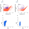
Flow Cytometry analysis of Extracellular Vesicles. Flow cytometry analysis of EVs from urine samples from a patient affected by low grade superficial papillary carcinoma of the urinary bladder (A) and from a healthy volunteer (B). EVs are identified as lipophilic cationic dye-positive (LCD) particles emitting on the allophycocyanin-APC-channel, and negative for phalloidin, emitting on the fluorescein isothiocyanate (FITC) channel. Dot density in the EV gate is higher in the urine of the patient. (C–D). Flow cytometry analysis of EVs from urine of patient affected with metastatic pheochromocytoma (C) relative to a healthy volunteer (D). EVs were analyzed for their scattered parameters on Forward Scatter (FSC)/Side Scatter (SSC) dot-plots. Number of events in the EV gate is higher in the urine of the patient.
Figure 3
Figure 3
Schematic representation of extracellular vesicle transcytosis through the blood brain barrier (BBB). The BBB is a complex physical barrier made up by endothelial cells in close association with pericytes and astrocytes. Extracellular vesicles circulating in peripheral blood first cross through transcytosis the tightly joined endothelial cells of the cerebral capillaries. Then, the vesicles interact with the next cell layers, pericytes and astrocytes. These sequential transfers imply mechanisms of selective recognition, trans-cellular transport and release. (modified from Servier Medical Art, licensed under a Creative Common Attribution 3.0 Unported License; http://smart.servier.com).

References

    1. Schopf J.W. Fossil evidence of Archaean life. Philos. Trans. R. Soc. Lond. B. Biol. Sci. 2006;361:869–885. doi: 10.1098/rstb.2006.1834. - DOI - PMC - PubMed
    1. Gribaldo S., Poole A.M., Daubin V., Forterre P., Brochier-Armanet C. The origin of eukaryotes and their relationship with the Archaea: Are we at a phylogenomic impasse? Nat. Rev. Microbiol. 2010;8:743–752. doi: 10.1038/nrmicro2426. - DOI - PubMed
    1. Reinhard C.T., Planavsky N.J., Olson S.L., Lyons T.W., Erwin D.H. Earth’s oxygen cycle and the evolution of animal life. Proc. Natl. Acad. Sci. USA. 2016;113:8933–8938. doi: 10.1073/pnas.1521544113. - DOI - PMC - PubMed
    1. Torday J.S., Rehan V.K. Cell-cell signaling drives the evolution of complex traits: Introduction-lung evo-devo. Integr. Comp. Biol. 2009;49:142–154. doi: 10.1093/icb/icp017. - DOI - PMC - PubMed
    1. Butterfield N.J. Modes of pre-Ediacaran multicellularity. Precambrian Res. 2009;173:201–211. doi: 10.1016/j.precamres.2009.01.008. - DOI

LinkOut - more resources