. 2020 Apr 5;10(4):688.
doi: 10.3390/nano10040688.

Bimetallic Core-Shell Nanoparticles of Gold and Silver via Bioinspired Polydopamine Layer as Surface-Enhanced Raman Spectroscopy (SERS) Platform

Affiliations

Bimetallic Core-Shell Nanoparticles of Gold and Silver via Bioinspired Polydopamine Layer as Surface-Enhanced Raman Spectroscopy (SERS) Platform

Asli Yilmaz et al. Nanomaterials (Basel). .

Abstract

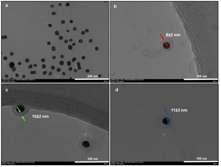
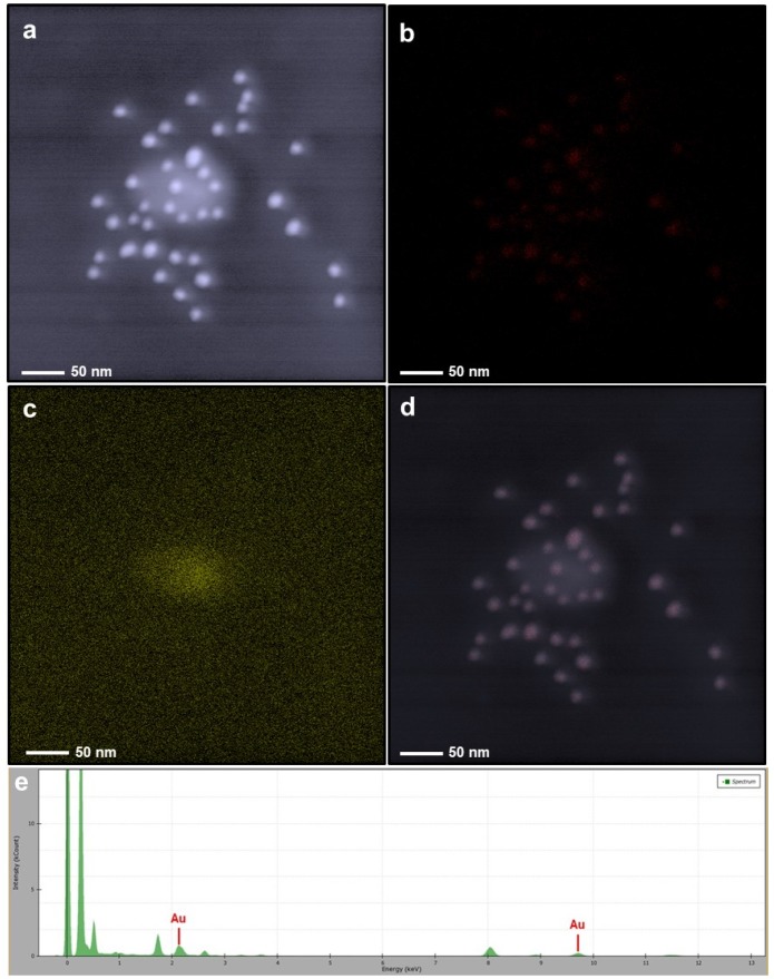
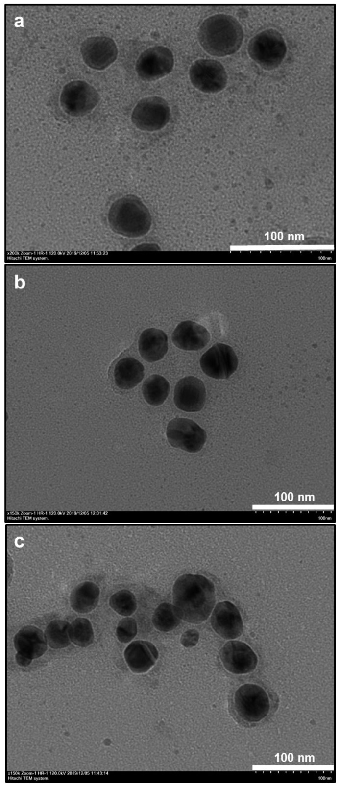
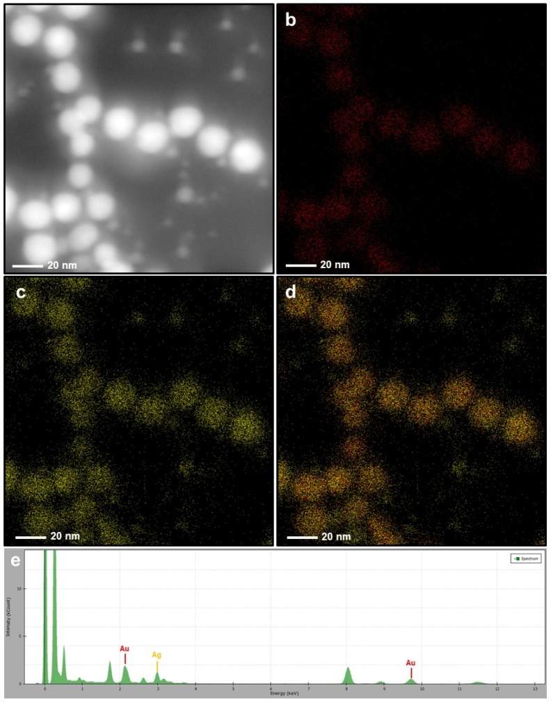
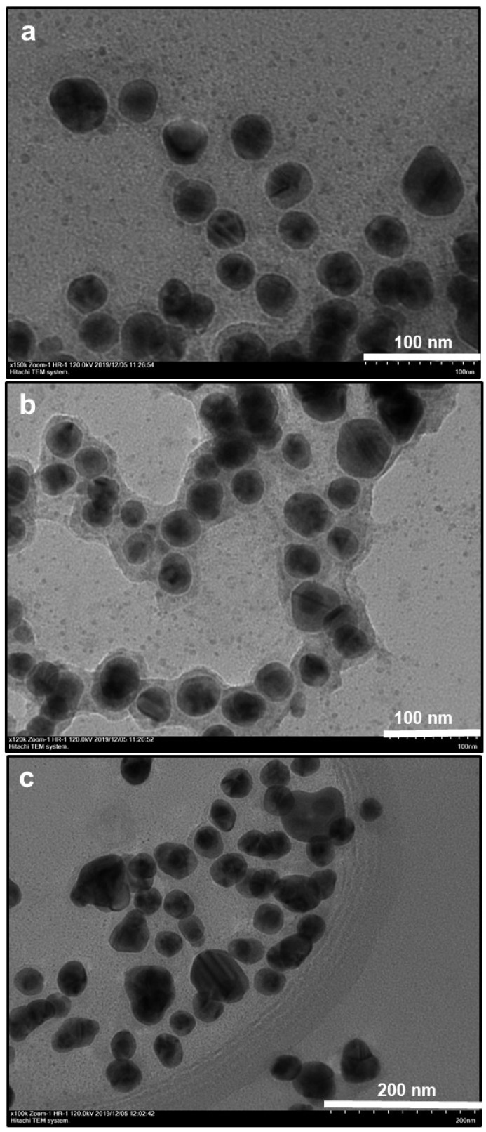
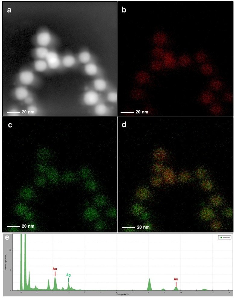
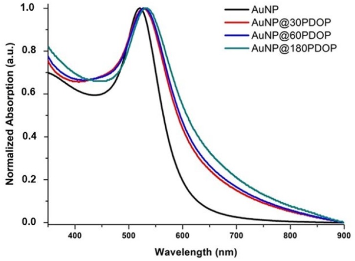
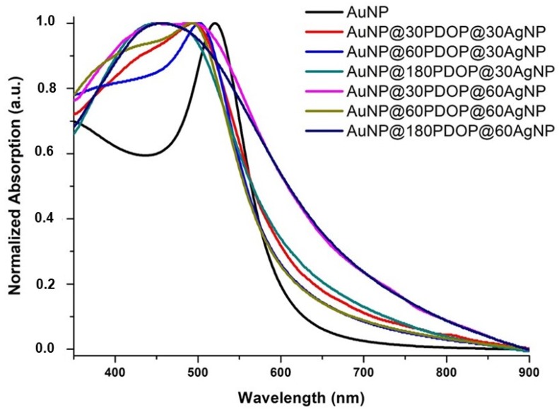
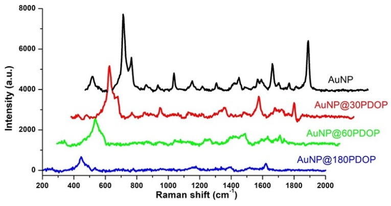
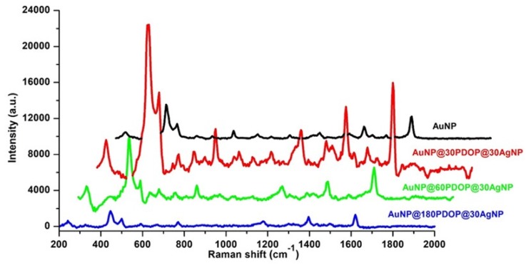
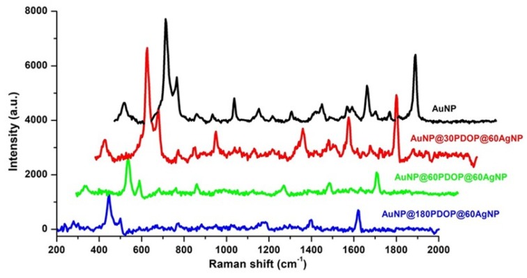
Despite numerous attempts to fabricate the core-shell nanoparticles, novel, simple, and low-cost approaches are still required to produce these efficient nanosystems. In this study, we propose the synthesis of bimetallic core-shell nanoparticles of gold (AuNP) and silver (AgNP) nanostructures via a bioinspired polydopamine (PDOP) layer and their employment as a surface-enhanced Raman spectroscopy (SERS) platform. Herein, the PDOP layer was used as an interface between nanostructures as well as stabilizing and reducing agents for the deposition of silver ions onto the AuNPs. UV-vis absorption spectra and electron microscope images confirmed the deposition of the silver ions and the formation of core-shell nanoparticles. SERS activity tests indicated that both the PDOP thickness and silver deposition time are the dominant parameters that determine the SERS performances of the proposed core-shell system. In comparison to bare AuNPs, more than three times higher SERS signal intensity was obtained with an enhancement factor of 3.5 × 10⁵.

Keywords: bimetallic core–shell nanoparticles; gold nanoparticles; polydopamine; surface-enhanced Raman spectroscopy (SERS).


Conflict of interest statement

The authors declare no conflict of interest.

Figures

Figure 1
Figure 1
TEM images ((a): bare AuNP, (b): AuNP@30PDOP, (c): AuNP@60PDOP, and (d): AuNP@180PDOP) of AuNP and AuNP@PDOP for different polydopamine (PDOP) deposition times (numbers indicate deposition times in min). AuNP@PDOP: PDOP-coated gold nanoparticles immersed into silver nitrate solution for different reduction times (10 mM AgNO3, 30, 60, and 180 min).
Figure 2
Figure 2
Elemental mapping and energy-dispersive X-ray spectroscopy (EDX) spectrum of the AuNP@30PDOP NP system. Electron micrograph region (a), distribution of gold elemental mapping (b), distribution of carbon elemental mapping (c), overlay of gold and carbon, and (d) relevant energy-dispersive X-ray spectroscopy spectrometer (EDAX) spectrum (e).
Figure 3
Figure 3
Effect of PDOP polymerization time (i.e., PDOP thickness) on core–shell NPs morphology for 30 min of silver reduction time. TEM images ((a): AuNP@30PDOP@30AgNP, (b): AuNP@60PDOP@30AgNP, and (c): AuNP@180PDOP@30AgNP) of a bimetallic core–shell NP system for different PDOP deposition times (numbers indicate deposition times in min).
Figure 4
Figure 4
Elemental mapping and EDAX spectrum of the AuNP@30PDOP@30AgNP system. Electron micrograph region (a), distribution of gold elemental mapping (b), distribution of silver elemental mapping (c), overlay of gold and silver (d), and relevant EDX spectrum (e).
Figure 5
Figure 5
Effect of PDOP polymerization time (i.e., PDOP thickness) on core–shell NPs morphology for 60 min of silver reduction time. TEM images ((a): AuNP@30PDOP@60AgNP, (b): AuNP@60PDOP@60AgNP, and (c): AuNP@180PDOP@60AgNP) of bimetallic core–shell NP system for different PDOP deposition times (numbers indicate deposition times in min).
Figure 6
Figure 6
Elemental mapping and EDAX spectrum of the AuNP@30PDOP@60AgNP system. Electron micrograph region (a), distribution of gold elemental mapping (b), distribution of silver elemental mapping (c), overlay of gold and silver (d), and relevant EDX spectrum (e).
Figure 7
Figure 7
UV-vis absorption spectra of AuNP and AuNP@PDOP for different PDOP deposition times (numbers indicate deposition times in min).
Figure 8
Figure 8
UV-vis absorption spectra of AuNP and AuNP@PDOP@AgNP for different PDOP and silver deposition times (numbers indicate deposition times in min).
Figure 9
Figure 9
Representative Raman measurements of AuNP and AuNP@PDOP for different PDOP deposition times (numbers indicate deposition times in min). The final concentration of methylene blue (MB) for Raman measurement is 10−5 M.
Figure 10
Figure 10
Effect of PDOP polymerization time (i.e., PDOP thickness) on SERS effect for 30 min of silver reduction time. Representative Raman measurements of the bimetallic core–shell NP system for different PDOP deposition times (numbers indicate deposition times in min). The final concentration of MB for Raman measurement is 10−5 M.
Figure 11
Figure 11
Effect of PDOP polymerization time (i.e., PDOP thickness) on SERS effect for 60 min of silver reduction time. Representative Raman measurements of the bimetallic core–shell NP system for different PDOP deposition times (numbers indicate deposition times in min). The final concentration of MB for Raman measurement is 10−5 M.

References

    1. Gawande M.B., Goswami A., Asefa T., Guo H., Biradar A.V., Peng D.-L., Zboril R., Varma R. Core–shell nanoparticles: Synthesis and applications in catalysis and electrocatalysis. Chem. Soc. Rev. 2015;44:7540–7590. doi: 10.1039/C5CS00343A. - DOI - PubMed
    1. Li J.-F., Zhang Y.-J., Ding S.-Y., Panneerselvam R., Tian Z.-Q. Core–shell nanoparticle-enhanced Raman spectroscopy. Chem. Rev. 2017;117:5002–5069. doi: 10.1021/acs.chemrev.6b00596. - DOI - PubMed
    1. Lim D.-K., Jeon K.-S., Hwang J.-H., Kim H., Kwon S., Suh Y.D., Nam J.-M. Highly uniform and reproducible surface-enhanced Raman scattering from DNA-tailorable nanoparticles with 1-nm interior gap. Nat. Nanotechnol. 2011;6:452. doi: 10.1038/nnano.2011.79. - DOI - PubMed
    1. Jiang H.-L., Akita T., Xu Q. A one-pot protocol for synthesis of non-noble metal-based core-shell nanoparticles under ambient conditions: Toward highly active and cost-effective catalysts for hydrolytic dehydrogenation of NH 3 BH 3. Chem. Commun. 2011;47:10999–11001. doi: 10.1039/c1cc13989d. - DOI - PubMed
    1. Caruso F., Spasova M., Salgueiriño-Maceira V., Liz-Marzán L. Multilayer assemblies of silica-encapsulated gold nanoparticles on decomposable colloid templates. Adv. Mater. 2001;13:1090–1094. doi: 10.1002/1521-4095(200107)13:14<1090::AID-ADMA1090>3.0.CO;2-H. - DOI

LinkOut - more resources