

Relationship Between Venules and Perivascular Spaces in Sporadic Small Vessel Diseases
- PMID: 32264759
- PMCID: PMC7185057
- DOI: 10.1161/STROKEAHA.120.029163
Relationship Between Venules and Perivascular Spaces in Sporadic Small Vessel Diseases
Abstract
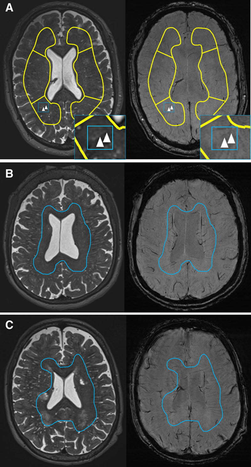
Background and Purpose- Perivascular spaces (PVS) around venules may help drain interstitial fluid from the brain. We examined relationships between suspected venules and PVS visible on brain magnetic resonance imaging. Methods- We developed a visual venular quantification method to examine the spatial relationship between venules and PVS. We recruited patients with lacunar stroke or minor nondisabling ischemic stroke and performed brain magnetic resonance imaging and retinal imaging. We quantified venules on gradient echo or susceptibility-weighted imaging and PVS on T2-weighted magnetic resonance imaging in the centrum semiovale and then determined overlap between venules and PVS. We assessed associations between venular count and patient demographic characteristics, vascular risk factors, small vessel disease features, retinal vessels, and venous sinus pulsatility. Results- Among 67 patients (69% men, 69.0±9.8 years), only 4.6% (range, 0%-18%) of venules overlapped with PVS. Total venular count increased with total centrum semiovale PVS count in 55 patients after accounting for venule-PVS overlap (β=0.468 [95% CI, 0.187-0.750]) and transverse sinus pulsatility (β=0.547 [95% CI, 0.309-0.786]) and adjusting for age, sex, and systolic blood pressure. Conclusions- Despite increases in both visible PVS and suspected venules, we found minimal spatial overlap between them in patients with sporadic small vessel disease, suggesting that most magnetic resonance imaging-visible centrum semiovale PVS are periarteriolar rather than perivenular.
Keywords: brain; humans; risk factors; small vessel disease; venular insufficiency, systemic; venules.
Figures
References
-
- Wardlaw JM, Smith EE, Biessels GJ, Cordonnier C, Fazekas F, Frayne R, et al. Standards for Reporting Vascular Changes on Neuroimaging (STRIVE v1) Neuroimaging standards for research into small vessel disease and its contribution to ageing and neurodegeneration. Lancet Neurol. 2013;12:822–838. doi: 10.1016/S1474-4422(13)70124-8. - PMC - PubMed
-
- Jochems ACC, Blair GW, Stringer MS, Thrippleton MJ, Clancy U, Chappell FM, et al. Methods to quantify and visualize venules using structural magnetic resonance imaging scans, 2000-2019 [dataset]. University of Edinburgh. Department of Neuroimaging Sciences. Centre for Clinical Brain Sciences. Date available: January 21, 2020. Edinburgh DataShare. doi: 10.7488/ds/2755. https://datashare.is.ed.ac.uk/handle/10283/3557.
Publication types
MeSH terms
Grants and funding
LinkOut - more resources
Full Text Sources
Research Materials
