

Abnormal development of cerebellar-striatal circuitry in Huntington disease
- PMID: 32265233
- PMCID: PMC7274924
- DOI: 10.1212/WNL.0000000000009364
Abnormal development of cerebellar-striatal circuitry in Huntington disease
Abstract
Objective: To test the hypothesis that the trajectory of functional connections over time of the striatum and the cerebellum differs between presymptomatic patients with the Huntington disease (HD) gene expansion (GE) and patients with a family history of HD but without the GE (GNE), we evaluated functional MRI data from the Kids-HD study.
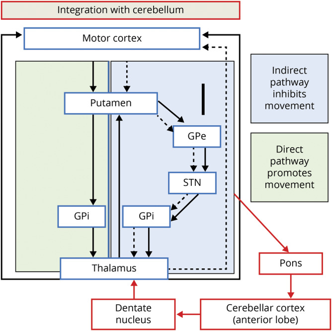
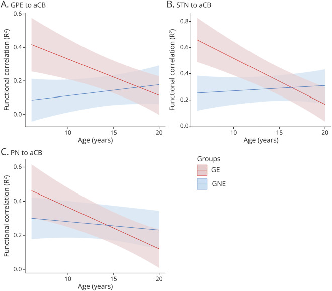
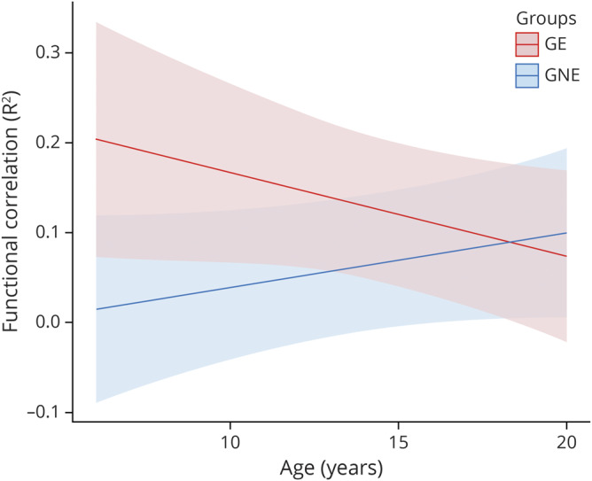
Methods: We utilized resting-state, functional MRI data from participants in the Kids-HD study between 6 and 18 years old. Participants were divided into GE (CAG 36-59) and GNE (CAG <36) groups. Seed-to-seed correlations were calculated among 4 regions that provide input signals to the anterior cerebellum: (1) dorsocaudal putamen, (2) globus pallidus externa, (3) subthalamic nucleus, and (4) pontine nuclei; and 2 regions that represented output from the cerebellum: the dentate nucleus to the (1) ventrolateral thalamus and (2) dorsocaudal putamen. Linear mixed effects regression models evaluated differences in developmental trajectories of these connections over time between groups.
Results: Four of the six striatal-cerebellum correlations showed significantly different trajectories between groups. All showed a pattern where in the early age ranges (6-12 years) there was hyperconnectivity in the GE compared to the GNE, with those trajectories showing linear decline in the latter half of the age range.
Conclusion: These results parallel previous findings showing striatal hypertrophy in children with GE as early as age 6. These findings support the notion of developmentally higher connectivity between the striatum and cerebellum early in the life of the child with HD GE, possibly setting the stage for cerebellar compensatory mechanisms.
© 2020 American Academy of Neurology.
Figures
References
-
- Norremolle A, Riess O, Epplen JT, Fenger K, Hasholt L, Sorensen SA. Trinucleotide repeat elongation in the Huntingtin gene in Huntington disease patients from 71 Danish families. Hum Mol Genet 1993;2:1475–1476. - PubMed
-
- Nasir J, Floresco SB, O'Kusky JR, et al. Targeted disruption of the Huntington's disease gene results in embryonic lethality and behavioral and morphological changes in heterozygotes. Cell 1995;81:811–823. - PubMed
-
- Zeitlin S, Liu JP, Chapman DL, Papaioannou VE, Efstratiadis A. Increased apoptosis and early embryonic lethality in mice nullizygous for the Huntington's disease gene homologue. Nat Genet 1995;11:155–163. - PubMed
Publication types
MeSH terms
Grants and funding
LinkOut - more resources
Full Text Sources
Medical