. 2020 May 15;295(20):6946-6957.
doi: 10.1074/jbc.RA120.012798. Epub 2020 Apr 7.

Arachidonate 12-lipoxygenase and 12-hydroxyeicosatetraenoic acid contribute to stromal aging-induced progression of pancreatic cancer

Affiliations

Arachidonate 12-lipoxygenase and 12-hydroxyeicosatetraenoic acid contribute to stromal aging-induced progression of pancreatic cancer

Ehab H Sarsour et al. J Biol Chem. .

Abstract

The incidence of pancreatic cancer increases with age, suggesting that chronological aging is a significant risk factor for this disease. Fibroblasts are the major nonmalignant cell type in the stroma of human pancreatic ductal adenocarcinoma (PDAC). In this study, we investigated whether the chronological aging of normal human fibroblasts (NHFs), a previously underappreciated area in pancreatic cancer research, influences the progression and therapeutic outcomes of PDAC. Results from experiments with murine xenografts and 2D and 3D co-cultures of NHFs and PDAC cells revealed that older NHFs stimulate proliferation of and confer resistance to radiation therapy of PDAC. MS-based metabolite analysis indicated that older NHFs have significantly increased arachidonic acid 12-lipoxygenase (ALOX12) expression and elevated levels of its mitogenic metabolite, 12-(S)-hydroxy-5,8,10,14-eicosatetraenoic acid (12-(S)-HETE) compared with their younger counterparts. In co-cultures with older rather than with younger NHFs, PDAC cells exhibited increases in mitogen-activated protein kinase signaling and cellular metabolism, as well as a lower oxidation state that correlated with their enhanced proliferation and resistance to radiation therapy. Expression of ALOX12 was found to be significantly lower in PDAC cell lines and tumor biopsies, suggesting that PDAC cells rely on a stromal supply of mitogens for their proliferative needs. Pharmacological (hydroxytyrosol) and molecular (siRNA) interventions of ALOX12 in older NHFs suppressed their ability to stimulate proliferation of PDAC cells. We conclude that chronological aging of NHFs contributes to PDAC progression and that ALOX12 and 12-(S)-HETE may be potential stromal targets for interventions that seek to halt progression and improve therapy outcomes.

Keywords: aging; arachidonic acid (AA) (ARA); cancer biology; cell proliferation; fibroblast; pancreatic cancer; stromal cell.


Conflict of interest statement

The authors declare that they have no conflicts of interest with the contents of this article

Figures

Figure 1.
Figure 1.
Older NHFs stimulate proliferation of MIA PaCa-2 human PDAC cells in vivo. A, 3D gel matrix-embedded co-cultures of CellTracker Green-labeled NHFs and luciferase expressing MIA PaCa-2-Luc cells were injected into the flank of mice, and imaged when palpable tumors were detected. n = 2. B, H&E and fluorescent microscopy detection of NHFs (yellow arrows) and MIA PaCa-2 cells (white arrows). C, flow cytometry analysis of cell suspensions prepared from excised tumors: CellTracker Green-positive (NHFs) and negative (MIA PaCa-2-Luc) cells. D, tumor volume of co-cultures of NHFs and MIA PaCa-2 cells; inset shows representative resected tumors. Asterisks represent significance; mean ± S.D.; n = 10, p < 0.05. E, Kaplan-Meyer plot of survival; n = 10, p < 0.05.
Figure 2.
Figure 2.
Chronological aging of NHFs influences progression and therapy of human PDAC cells in 3D co-cultures. A, pseudo-color scale of photons s−1 emitted from individual 3D spheres of co-cultures of NHFs and MIA PaCa-2-luc cells. B, photon counts of MIA PaCa-2-luc cells in co-cultures; inset shows H&E staining of sliced spheres: fibroblasts (black arrows), cancer cells (white arrows). Photon counts of (C) MIA PaCa-2-Luc and (D) PANC-1-Luc cells in 3D co-cultures of NHFs post-radiation (2 Gy × 5 days). Asterisks represent significance; n = 3, p < 0.05.
Figure 3.
Figure 3.
Older NHFs stimulate proliferation of PDAC cells in 2D co-cultures. A, microscopy pictures of 5-day co-cultures of NHFs and CellTracker Green-labeled MIA PaCa-2 cells. B, growth of CellTracker Green-labeled MIA PaCa-2 cells in co-cultures of NHFs. C, flow cytometry analysis of MIA PaCa-2 cells in co-cultures of NHFs. D, representative flow cytometry histograms showing distributions of NHFs and MIA PaCa-2 cells in co-cultures. E, percentage of PDAC cells in co-cultures of NHFs from donors of different ages. Asterisks represent significance; n = 3, p < 0.05.
Figure 4.
Figure 4.
Resistance of MIA PaCa-2 cells to radiation treatments in 2D co-cultures of old compared with young NHFs. CellTracker Green-labeled MIA PaCa-2 cells were layered over confluent cultures of NHFs from 3-day- and 12- and 61-year-old healthy donors. At the end of 5 days of co-cultures, cells were irradiated with 0, 2, and 4 Gy of ionizing radiation. Flow cytometry was used to identify and quantitate CellTracker Green-positive (MIA PaCa-2) and negative (NHFs) cells. Proliferation of MIA PaCa-2 cells was assessed from the decreases in CellTracker Green fluorescence per cell. A, percent of MIA PaCa-2 cells in control and irradiated co-cultures of NHFs; B, percent of MIA PaCa-2 cell death in control and irradiated co-cultures; n = 3, p < 0.05.
Figure 5.
Figure 5.
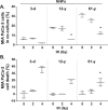
An age-related increase in ALOX12 expression and 12-(S)-HETE is associated with old NHFs-induced stimulation of PDAC cells. A, mass spectrometry analysis of lipid metabolites in conditioned media of 3-day-old and 61-year-old NHFs. Fold-change was calculated relative to metabolites in 3-day-old NHFs. B, RT-qPCR analysis of arachidonic acid-metabolizing enzymes; inset shows immunoblot analysis of ALOX12. An ELISA-based assay was used to quantitatively measure 12-(S)-HETE levels in conditioned media collected from: C, quiescent cultures of NHFs from donors of different ages and D, 3-day-old NHFs that were aged in culture. RT-qPCR analysis: ALOX12 expression in: E, PDAC cell lines obtained from ATCC and F, newly generated PDAC cell lines from surgically resected and de-identified human PDAC tissues. G, RT-qPCR analysis of GPR31 mRNA expression. H, proliferation of MIA PaCa-2 cells in the presence of exogenously added 12-(S)-HETE. Asterisks represent significance; n = 3, p < 0.05.
Figure 6.
Figure 6.
Metabolic and proliferative signaling pathways are enhanced in PDAC cells co-cultured with older NHFs. RayBiotech MAPK Pathway Phosphorylation Arrays were used to analyze the phosphorylation status of signaling proteins of MIA PaCa-2 cells sorted from co-cultures of NHFs. Visualization and quantification of results were performed using a Typhoon 7000 phosphorimager (GE Healthcare) and NIH ImageJ software. Left panels, images of the original blots and heat maps; right panel, quantitative results. Percent increase in phosphoproteins of 61-year-old NHFs was calculated relative to 3-day-old NHFs.
Figure 7.
Figure 7.
Increases in metabolism combined with a lower oxidation status of PDAC cells co-cultured with old compared with young NHFs. A, flow cytometry sorted MIA PaCa-2 cells from co-cultures of CellTracker Green-labeled NHFs were used for analysis of metabolites (UIOWA HCCC Metabolomics Core). Results were normalized to cell mass and fold-change calculated relative to metabolites of MIA PaCa-2 cells co-cultured with 3-day-old NHFs. B and C, flow cytometry analysis of DHE-oxidation in co-cultures of CellTracker Green-labeled NHFs and MIA PaCa-2 cells. Asterisk represents significance compared with 3-day-old NHFs; n = 3, p < 0.05.
Figure 8.
Figure 8.
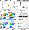
Hydroxytyrosol and siALOX12 treatments suppress older NHFs induced stimulation of PDAC proliferation. A, 12-(S)-HETE levels in conditioned media from 10-day quiescent cultures of control and 100 μm hydroxytyrosol-treated NHFs. B, flow cytometry analysis of the percentage of MIA PaCa-2 cells in co-cultures of CellTracker Green-labeled control and hydroxytyrosol-treated NHFs. C, plating efficiency of MIA PaCa-2 cells cultured in conditioned media from control and hydroxytyrosol-treated NHFs. Asterisks represent statistical significance; n = 3, p < 0.05. D, upper panel: immunoblot analysis of ALOX12 protein levels in control, scrambled siRNA, and siALOX12 treated 61-year-old NHFs. Lower panel: flow cytometry analysis of the percentage of MIA PaCa-2 cells in co-cultures of NHFs.
Figure 9.
Figure 9.
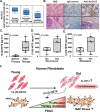
ALOX12 and 12-(S)-HETE are potential stromal-aging biomarkers for PDAC progression and therapy response. A, Oncomine data analysis of ALOX12 expression in pancreas cancer (37). B, immunohistochemistry analysis of ALOX12 in paraffin-embedded and de-identified resected PDAC tissues: black arrows, PDAC cells; yellow arrows, nonmalignant stromal cells. C, 12-(S)-HETE levels in de-identified plasma samples from 14 PDAC patients. Results were evaluated for incidence of (D) recurrence and (E) metastasis. Asterisks represent significance, p < 0.05, measured by paired t test. F, an illustration showing increases in ALOX12 expression and 12-(S)-HETE levels during chronological aging of NHFs promoting PDAC progression by enhancing MAPK signaling pathways and metabolism. It is hypothesized that increases in metabolism combined with a reducing status confer resistance of PDAC cells to therapy in an aging-stromal environment.

References

    1. Hidalgo M. (2010) Pancreatic cancer. N. Engl. J. Med. 362, 1605–1617 10.1056/NEJMra0901557 - DOI - PubMed
    1. Yip D., Karapetis C., Strickland A., Steer C. B., and Goldstein D. (2006) Chemotherapy and radiotherapy for inoperable advanced pancreatic cancer. Cochrane Database Syst. Rev. 19, CD002093 - PubMed
    1. Tang D. G., Bhatia B., Tang S., and Schneider-Broussard R. (2007) 15-Lipoxygenase 2 (15-LOX2) is a functional tumor suppressor that regulates human prostate epithelial cell differentiation, senescence, and growth (size). Prostaglandins Other Lipid Mediat. 82, 135–146 10.1016/j.prostaglandins.2006.05.022 - DOI - PubMed
    1. Fang H., and Declerck Y. A. (2013) Targeting the tumor microenvironment: from understanding pathways to effective clinical trials. Cancer Res. 73, 4965–4977 10.1158/0008-5472.CAN-13-0661 - DOI - PMC - PubMed
    1. Kadaba R., Birke H., Wang J., Hooper S., Andl C. D., Di Maggio F., Soylu E., Ghallab M., Bor D., Froeling F. E., Bhattacharya S., Rustgi A. K., Sahai E., Chelala C., Sasieni P., and Kocher H. M. (2013) Imbalance of desmoplastic stromal cell numbers drives aggressive cancer processes. J. Pathol. 230, 107–117 10.1002/path.4172 - DOI - PMC - PubMed

Publication types

MeSH terms

Substances