Comparative Study
. 2020 Jun;61(6):884-895.
doi: 10.1194/jlr.RA120000671. Epub 2020 Apr 7.

Comparative profiling and comprehensive quantification of stratum corneum ceramides in humans and mice by LC/MS/MS

Affiliations
Comparative Study

Comparative profiling and comprehensive quantification of stratum corneum ceramides in humans and mice by LC/MS/MS

Momoko Kawana et al. J Lipid Res. 2020 Jun.

Abstract

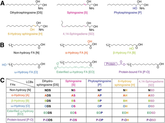
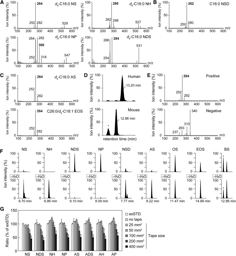
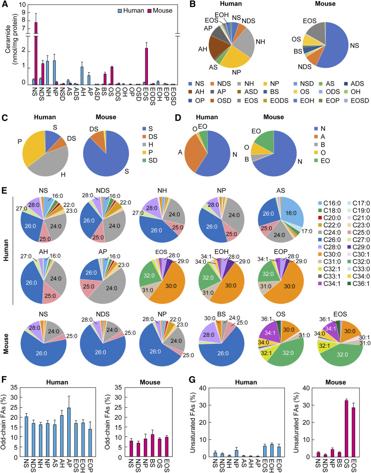
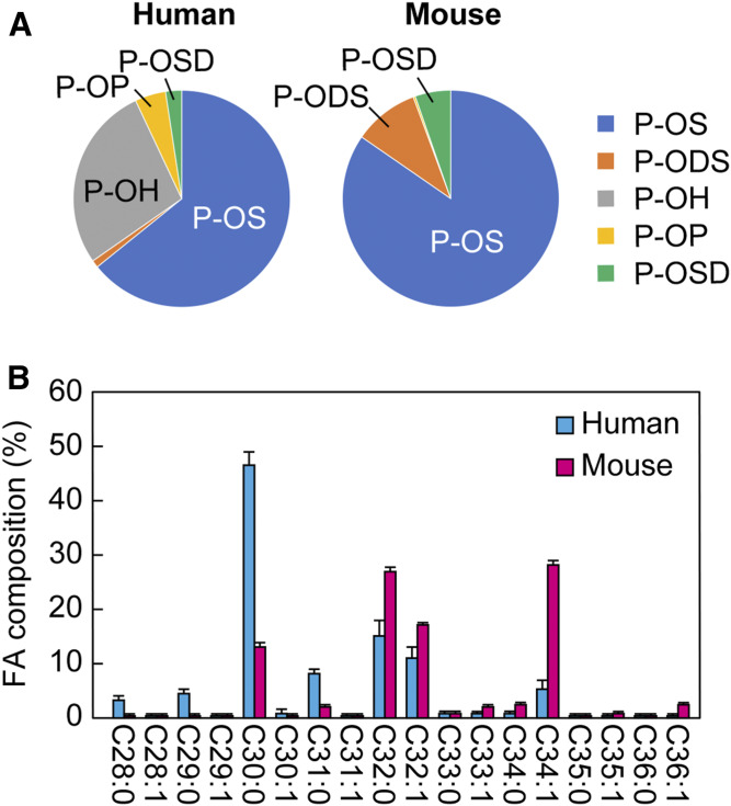
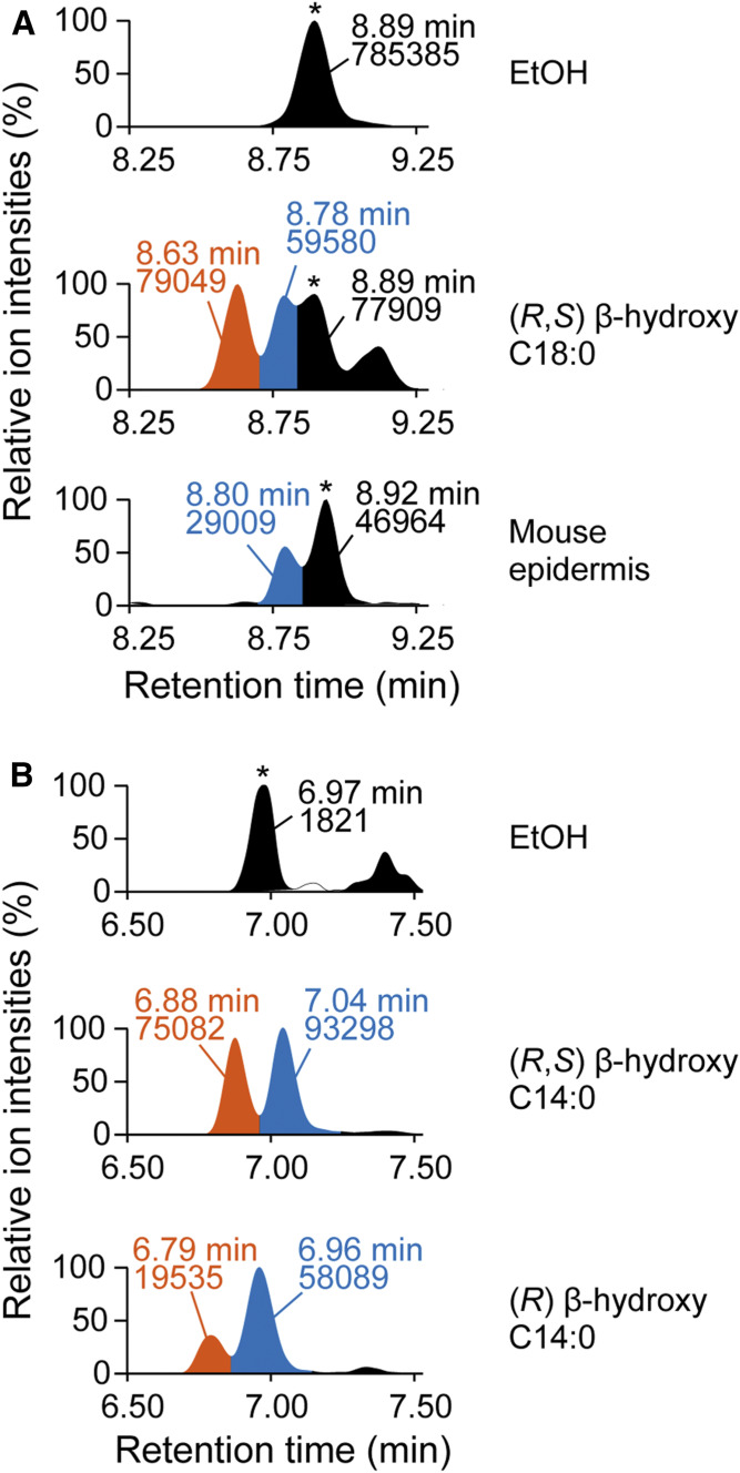
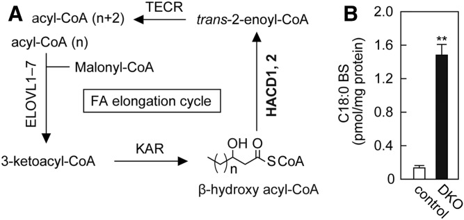
Ceramides are the predominant lipids in the stratum corneum (SC) and are crucial components for normal skin barrier function. Although the composition of various ceramide classes in the human SC has been reported, that in mice is still unknown, despite mice being widely used as animal models of skin barrier function. Here, we performed LC/MS/MS analyses using recently available ceramide class standards to measure 25 classes of free ceramides and 5 classes of protein-bound ceramides from human and mouse SC. Phytosphingosine- and 6-hydroxy sphingosine-type ceramides, which both contain an additional hydroxyl group, were abundant in the human SC (35% and 45% of total ceramides, respectively). In contrast, in mice, phytosph-ingosine- and 6-hydroxy sphingosine-type ceramides were present at ∼1% and undetectable levels, respectively, and sphingosine-type ceramides accounted for ∼90%. In humans, ceramides containing α-hydroxy FA were abundant, whereas ceramides containing β-hydroxy or ω-hydroxy FA were abundant in mice. The hydroxylated β-carbon in β-hydroxy ceramides was in the (R) configuration. Genetic knockout of β-hydroxy acyl-CoA dehydratases in HAP1 cells increased β-hydroxy ceramide levels, suggesting that β-hydroxy acyl-CoA, an FA-elongation cycle intermediate in the ER, is a substrate for β-hydroxy ceramide synthesis. We anticipate that our methods and findings will help to elucidate the role of each ceramide class in skin barrier formation and in the pathogenesis of skin disorders.

Keywords: epidermis; fatty acid; lipidomics; mass spectrometry; skin barrier; sphingolipids.


Conflict of interest statement

The authors declare that they have no conflicts of interest with the contents of this article

Figures

Fig. 1.
Fig. 1.
Structures and nomenclature for ceramide classes in mammals. A: Structures of mammalian LCBs. B: FAs constituting mammalian ceramides. C: Nomenclature for 25 free ceramide classes and five P-O ceramide classes. Each ceramide class is represented by a combination of the abbreviations corresponding to its FA and LCB structure.
Fig. 2.
Fig. 2.
Optimization of SC ceramide analysis using LC/MS/MS. A–C: Product ion scanning was performed using ceramide standards in the positive ion mode (A: d9-C16:0 NS, d9-C16:0 NDS, d9-C16:0 NH, and d9-C16:0 NP; B: C16:0 NSD; and C: d9-C16:0 AS and C26:0/d9-C18:1 EOS). The m/z values of each precursor ion were set as follows: d9-C16:0 NS, 547.5; d9-C16:0 NDS, 549.5; d9-C16:0 NH, 563.5; d9-C16:0 NP, 565.5; C16:0 NSD, 536.5; d9-C16:0 AS, 563.5; and C26:0/d9-C18:1 EOS, 967.9. Fragment ions were detected in the scanning range m/z 100–600. The values shown in bold represent the m/z of a fragment ion specific for each ceramide class. C16:0 NSD was synthesized by incubating 1 µM SD and 1 µM C16:0-CoA with membrane fractions (50 µg) prepared from HEK 293T cells overexpressing CERS5 at 37°C for 1 h (B). D: Lipids were extracted from the human SC and mouse epidermis and subjected to LC/MS/MS analysis using the MRM setting to detect C26:0 AS. *Unidentified peak. E: Product ion scanning of C26:0 BS was performed in the positive ion mode (upper) and negative ion mode (lower). F: MRM analyses were performed by setting the m/z values of [M + H]+ (upper panel) and [M–H2O + H]+ (lower panel) at Q1 and the m/z values of a fragment ion specific for each LCB at Q3. The values below the horizontal axis represent the retention time of each peak. *Unidentified peak. G: Each d9 ceramide standard was added to the extract from the indicated size of tape piece (the final volumes of all samples were adjusted to 100 µl) and detected by LC/MS/MS. Values are the detection rate compared with the external standards (exSTD) and represent means ± SDs (n = 3; *P < 0.05 and **P < 0.01; Dunnett’s test).
Fig. 3.
Fig. 3.
Ceramide profiles in human and mouse SC. SC samples were collected by tape stripping from humans (aged 20–50 years; n = 19) and mice (on the first day after birth; n = 3). Lipids were extracted from the tapes and subjected to LC/MS/MS analyses to quantify ceramides. A: Amount of each ceramide class. Values are the sum of the ceramide species containing each FA chain length (nonhydroxy ceramides, α-hydroxy ceramides, and BS: C14–C36; ω-hydroxy and EO ceramides: C26–36) and represent means ± SDs. B: The ratio of each ceramide class to total ceramides. C: The proportion of ceramide classes containing a common LCB. D: The ratio of ceramide classes containing a common type of FA. E: The FA composition of ceramide classes representing more than 1% of the total ceramides in human and mouse SC. F: The ratio of odd-chain FAs in each ceramide class. Values represent means ± SDs. G: The ratio of unsaturated FAs in each ceramide class. Values represent means ± SDs.
Fig. 4.
Fig. 4.
P-O ceramide profiles in human and mouse SC. P-O ceramides were extracted from the SC samples collected by tape stripping from humans (aged 20–50 years; n = 19) and mice (on the first day after birth; n = 3) and quantified by LC/MS/MS analyses. A: The ratio of each P-O ceramide class to the total P-O ceramides. B: FA composition of P-OS. Values represent means ± SDs.
Fig. 5.
Fig. 5.
(R) configuration of hydroxylated β-carbon in BS. A: Membrane fractions (20 μg) prepared from HEK 293T cells overexpressing CERS1 were incubated with EtOH or 5 µM (R, S) β-hydroxy C18:0 FA and 5 μM d7 sphingosine at 37°C for 1 h to produce C18:0 BS. Lipids were extracted and subjected to LC/MS/MS analysis to detect C18:0 BS containing d7-labeled sphingosine [upper, EtOH; middle, (R, S) β-hydroxy C18:0 FA; and lower, C18:0 BS in mouse epidermis]. *Unidentified peak. B: Membrane fractions (20 μg) prepared from HEK 293T cells overexpressing CERS5 were incubated with EtOH, 5 µM (R, S) β-hydroxy C14:0 FA, or (R) β-hydroxy C14:0 FA and 5 μM d7 sphingosine at 37°C for 1 h. Lipids were extracted and subjected to LC/MS/MS analysis to detect C14:0 BS containing d7-labeled sphingosine [upper, EtOH; middle, (R,S) β-hydroxy C14:0 FA; and lower, (R) β-hydroxy C14:0 FA]. *Unidentified peak. The vertical axis of each chromatogram shows relative intensity. The values represented in the chromatogram indicate retention time (upper) and peak area (lower). EtOH, ethanol.
Fig. 6.
Fig. 6.
Increase in BS levels due to the accumulation of β-hydroxy acyl-CoAs, which are intermediates in the FA elongation cycle. A: The FA elongation cycle localized in the ER produces acyl-CoAs having ≥C18 chain lengths. In each cycle, the chain length of the substrate acyl-CoA is elongated by two carbon chains through a four-step reaction. The enzymes involved in each reaction step and intermediates are depicted. β-Hydroxy acyl-CoA dehydratases HACD1 and HACD2 convert β-hydroxy acyl-CoAs to trans-2-enoyl-CoAs. B: Lipids were extracted from control and HACD1 HACD2 DKO HAP1 cells, and C18:0 BS was quantified by LC/MS/MS. Values represent means ± SDs (n = 3; **P < 0.01; two-tailed Student’s t-test).

References

    1. Traupe H., Fischer J., and Oji V.. 2014. Nonsyndromic types of ichthyoses – an update. J. Dtsch. Dermatol. Ges. 12: 109–121. - PubMed
    1. Borodzicz S., Rudnicka L., Mirowska-Guzel D., and Cudnoch-Jedrzejewska A.. 2016. The role of epidermal sphingolipids in dermatologic diseases. Lipids Health Dis. 15: 13. - PMC - PubMed
    1. Goleva E., Berdyshev E., and Leung D. Y.. 2019. Epithelial barrier repair and prevention of allergy. J. Clin. Invest. 129: 1463–1474. - PMC - PubMed
    1. Yardley H. J., and Summerly R.. 1981. Lipid composition and metabolism in normal and diseased epidermis. Pharmacol. Ther. 13: 357–383. - PubMed
    1. Masukawa Y., Narita H., Shimizu E., Kondo N., Sugai Y., Oba T., Homma R., Ishikawa J., Takagi Y., Kitahara T., et al. 2008. Characterization of overall ceramide species in human stratum corneum. J. Lipid Res. 49: 1466–1476. - PubMed

Publication types