. 2020 Apr 7;11(2):e00305-20.
doi: 10.1128/mBio.00305-20.

Nonproteolytic K29-Linked Ubiquitination of the PB2 Replication Protein of Influenza A Viruses by Proviral Cullin 4-Based E3 Ligases

Affiliations

Nonproteolytic K29-Linked Ubiquitination of the PB2 Replication Protein of Influenza A Viruses by Proviral Cullin 4-Based E3 Ligases

Marwah Karim et al. mBio. .

Abstract

The multifunctional nature of viral proteins is essentially driven by posttranslational modifications (PTMs) and is key for the successful outcome of infection. For influenza A viruses (IAVs), a composite pattern of PTMs regulates the activity of viral proteins. However, almost none are known that target the PB2 replication protein, except for inducing its degradation. We show here that PB2 undergoes a nonproteolytic ubiquitination during infection. We identified E3 ubiquitin ligases catalyzing this ubiquitination as two multicomponent RING-E3 ligases based on cullin 4 (CRL4s), which are both contributing to the levels of ubiquitinated forms of PB2 in infected cells. The CRL4 E3 ligase activity is required for the normal progression of the viral cycle and for maximal virion production, indicating that the CRL4s mediate a ubiquitin signaling that promotes infection. The CRL4s are recruiting PB2 through an unconventional bimodal interaction with both the DDB1 adaptor and DCAF substrate receptors. While able to bind to PB2 when engaged in the viral polymerase complex, the CRL4 factors do not alter transcription and replication of the viral segments during infection. CRL4 ligases catalyze different patterns of lysine ubiquitination on PB2. Recombinant viruses mutated in the targeted lysines showed attenuated viral production, suggesting that CRL4-mediated ubiquitination of PB2 contributes to IAV infection. We identified K29-linked ubiquitin chains as main components of the nonproteolytic PB2 ubiquitination mediated by the CRL4s, providing the first example of the role of this atypical ubiquitin linkage in the regulation of a viral infection.IMPORTANCE Successful infection by influenza A virus, a pathogen of major public health importance, involves fine regulation of the multiple functions of the viral proteins, which often relies on post-translational modifications (PTMs). The PB2 protein of influenza A viruses is essential for viral replication and a key determinant of host range. While PTMs of PB2 inducing its degradation have been identified, here we show that PB2 undergoes a regulating PTM signaling detected during infection, based on an atypical K29-linked ubiquitination and mediated by two multicomponent E3 ubiquitin ligases. Recombinant viruses impaired for CRL4-mediated ubiquitination are attenuated, indicating that ubiquitination of PB2 is necessary for an optimal influenza A virus infection. The CRL4 E3 ligases are required for normal viral cycle progression and for maximal virion production. Consequently, they represent potential candidate host factors for antiviral targets.

Keywords: K29-linked ubiquitination; PB2 replication protein; cullin-based E3 ligases; influenza virus; nonproteolytic ubiquitination; post-translational modification; ubiquitination.


Figures

FIG 1
FIG 1
Involvement of CRL4 factors in IAV infection. (A) A549 cells were transfected with siRNA nontarget (NT) or siRNA targeting CRL4 factors (si target) for 48 h and then infected at an MOI of 0.0001 PFU/cell (H1N1WSN) or 0.001 PFU/cell (H1N1pdm09 and H3N2). Viral titers were determined by plaque-forming assay at the indicated time points. Statistical significances are given in Table S1. (B) A549 cells transfected with the indicated siRNA for 48 h were infected with H1N1pdm09 at an MOI of 3 PFU/cell. Total cell lysates were prepared at the indicated time postinfection and analyzed by Western blotting using the indicated antibodies. The relative amounts of viral proteins in siRNA-treated samples compared to the corresponding siRNA nontarget (NT) samples are indicated. (C) A549 cells transfected with the indicated siRNA for 48 h were infected with H1N1WSN at an MOI of 5 PFU/cell. At 6 h postinfection, cells were fixed, permeabilized, and stained with an anti-NP antibody (green) and with Hoechst 33342 (blue). Representative images of NP localization are shown. Quantification of the NP labeling is provided in Fig. S2C. (D) HEK293T cells were noninfected (NI) or infected with H1N1WSN (left) or H1N1WSN-PB2-Strep (right) virus at an MOI of 3 for 6 h. Cell lysate was subjected to anti-PB2 antibody and IgG (left) or to Strep-Tactin pulldown (right). Proteins in the pulled fractions and in 1/10 of whole-cell extract (inputs) were assessed in an immunoblot assay as indicated. (E) HEK293 cells stably expressing Strep-DDB1, Strep-DCAFs (D12L1 for DCAF12L1 or D11 for DCAF11), or Strep-mCherry (control) were infected with H1N1WSN at an MOI of 3 for 6 h and subjected to Strep-Tactin pulldown. The PB2 protein copulled with the Strep-CRL4 factors was detected using anti-PB2 antibody. One-fourth of whole-cell extract was used to detect the Strep-CRL4 factors in the input since they were undetected in a 1/10 fraction, indicating a low level of Strep fusion expression. The asterisk indicates an aspecific band.
FIG 2
FIG 2
Interaction between the CRL4 complexes and the viral polymerase. (A) Detection of DDB1-PB2-DCAF triple complexes. A schematic of the triple-complex pulldown is given where Strep tag is represented by a cyan sphere and the Strep-Tactin beads are represented by blue spheres. The Gluc2-PB2, Gluc1-DCAFs (either DCAF12L1 or DCAF11), and Strep-DDB1 or Strep-empty expression plasmids were cotransfected in HEK293T cells. At 24 h posttransfection, cell lysates were subjected to pulldown using Strep-Tactin Sepharose beads. The Gaussia luciferase activity retained on the beads was expressed as the proportion of Gaussia activity in the whole-cell lysate (% of input). The Gaussia activity in the whole-cell lysate, representing the level of interacting protein pair, is given in Fig. S3A. Data represent means ± SD for three experiments, and significance was measured using a two-tailed unpaired t test (nonsignificant [ns], P > 0.05; *, P ≤ 0.05; **, P ≤ 0.01; ***, P ≤ 0.001; ****, P ≤ 0.0001). (B) Interaction domains of PB2 with the CRL4 factors. HEK293T cells were transfected with Strep-DDB1, Strep-DCAF12L1, or Strep-DCAF11 together with 3×Flag-PB2 full-length (FL), N-terminal (Nt), or C-terminal (Ct) domains or with 3×Flag-empty expression plasmids. At 24 h posttransfection, cells were lysed and Strep-Tactin pulldown was performed. Proteins in the pulled fraction and in 1/10 of whole-cell extract (inputs) were analyzed by Western blotting as shown. (C) Interaction of CRL4 factors with the RNA-dependent RNA polymerase (RdRP). A schematic of the quaternary-complex pulldown is given. HEK293T cells were cotransfected with Strep-DDB1, Strep-DCAF12L1, Strep-DCAF11, or Strep-empty vector along with either 3×Flag-PB2 or 3×Flag-empty and with Gluc1-PA and PB1-Gluc2. At 24 h posttransfection, cells were lysed and subjected to pulldown using Strep-Tactin Sepharose beads. The retention of Gaussia activity on the beads was expressed as the proportion of the Gaussia activity in the whole-cell lysate (% of input). The Gaussia activity in the whole-cell lysate before pulldown, representing the level of interacting PB1/PA dimer, is given in Fig. S3C. Data represent means ± SD for three independent experiments, and the significance was measured using one-way ANOVA.
FIG 3
FIG 3
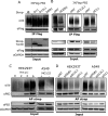
CRL4 E3 ligase complexes mediate PB2 ubiquitination. (A) HEK293T cells were transiently transfected with the indicated expression plasmids. At 36 h posttransfection, cells were treated with MG132 (10 μM) for 4 h. Anti-Flag immunoprecipitation (IP) was performed from cell lysates prepared under strong denaturing conditions (2% SDS), and the ubiquitinated forms of PB2 were detected using antiubiquitin antibody, followed by an anti-Flag immunoblot assay to detect PB2. Expression of Flag-PB2 and the Strep fusion proteins was monitored in cell lysate (Input). A dashed line marks that a lane from the initial membrane has been removed (not shown here). (B) HEK293 cells stably expressing Strep-DDB1, Strep-DCAF12L1 (D12L1), or Strep-DCAF11 (D11) were transfected with the indicated siRNA and 24 h later transfected with 3×Flag-PB2 for 36 h and treated with MG132 (10 μM) for 4 h. Cell lysate was processed for anti-Flag immunoprecipitation and immunoblotting as shown in panel A. (C) HEK293T cells were infected with H1N1WSN-Strep at an MOI of 3 for different times and treated with MG132 (10 μM) for 4 h before cell lysis (left). Similarly, A549 cells were infected with H1N1WSN-Strep at an MOI of 3 for 8 h and treated or not with 10 μM MG132 4 h before cell lysis (right). Strep-PB2 protein was pulled using Strep-Tactin Sepharose beads, and antiubiquitin and anti-PB2 immunoblotting assays were performed. (D) HEK293T and A549 cells transfected with the indicated siRNA for 48 h were infected with H1N1WSN-Strep at an MOI of 3 for 6 and 8 h, respectively, and treated with MG132 (10 μM) for 4 h before cell lysis. PB2-Strep pulldown and immunoblot assays were performed as described for panel C.
FIG 4
FIG 4
CRL4D11 and CRL4D12L1 mediate the nondegradative ubiquitination of PB2. (A) HEK293T cells were cotransfected with expression plasmids for 3×FLAG-PB2 and Strep-DDB1, Strep-DCAF12L1 (D12L1), Strep-DCAF11 (D11), or Strep-empty for 36 h and then treated with MG132 (+) for 4 h before lysis where indicated. Whole-cell lysates were prepared using Laemmli buffer and immunoblotted using indicated antibodies. (B) HEK293T cells were cotransfected with Strep-DDB1, Strep-DCAF12L1, or Strep-DCAF11 and with indicated HA-tagged ubiquitin mutants. At 36 h posttransfection, cells were lysed under strong denaturing conditions and subjected to Flag immunoprecipitation, followed by Western blotting with indicated antibodies.
FIG 5
FIG 5
CRL4s mediate different patterns of PB2 ubiquitination that are involved in infection. (A) HEK293 cells stably expressing Strep-DCAF12L1 or Strep-DCAF11 were cotransfected with the indicated PB2 mutants fused with 3×Flag tag together with HA-ubiquitin. At 48 h posttransfection, cell lysates were subjected to Flag pulldown and analyzed by immunoblotting with the indicated antibodies. Expression of Flag-PB2 and Strep fusions was monitored in cell lysate (Input). (B) A549 cells infected at an MOI of 0.001 with recombinant H1N1pdm09 viruses, either wild type or mutated in the PB2 lysines targeted by each of the CRL4 E3 ligases as indicated. Viral titers were determined in triplicates by plaque-forming assay at the indicated time points, and significance was measured using one-way ANOVA (ns, P > 0.05; *, P ≤ 0.05; **, P ≤ 0.01; ***, P ≤ 0.001).

References

    1. Swatek KN, Komander D. 2016. Ubiquitin modifications. Cell Res 26:399–422. doi:10.1038/cr.2016.39. - DOI - PMC - PubMed
    1. Chen ZJ, Sun LJ. 2009. Nonproteolytic functions of ubiquitin in cell signaling. Mol Cell 33:275–286. doi:10.1016/j.molcel.2009.01.014. - DOI - PubMed
    1. Pickart CM, Fushman D. 2004. Polyubiquitin chains: polymeric protein signals. Curr Opin Chem Biol 8:610–616. doi:10.1016/j.cbpa.2004.09.009. - DOI - PubMed
    1. Dwane L, Gallagher WM, Ní Chonghaile T, O’Connor DP. 2017. The emerging role of non-traditional ubiquitination in oncogenic pathways. J Biol Chem 292:3543–3551. doi:10.1074/jbc.R116.755694. - DOI - PMC - PubMed
    1. Ohtake F, Tsuchiya H. 2017. The emerging complexity of ubiquitin architecture. J Biochem 161:125–133. doi:10.1093/jb/mvw088. - DOI - PubMed

Publication types

LinkOut - more resources