Review
. 2020 Mar 20:10:112.
doi: 10.3389/fcimb.2020.00112. eCollection 2020.

Malassezia-Associated Skin Diseases, the Use of Diagnostics and Treatment

Affiliations
Review

Malassezia-Associated Skin Diseases, the Use of Diagnostics and Treatment

Ditte M L Saunte et al. Front Cell Infect Microbiol. .

Abstract

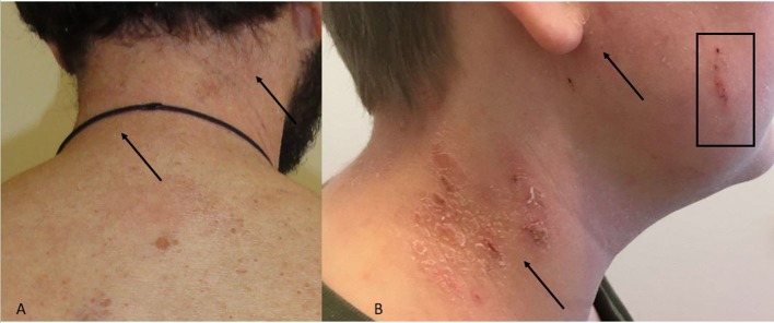
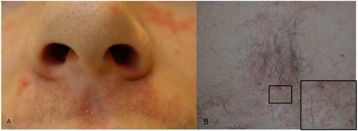
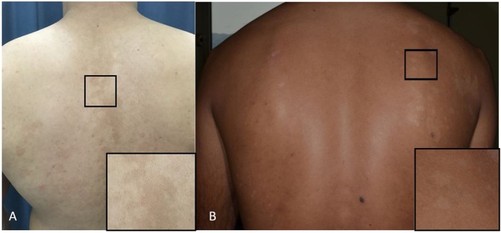
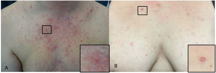
Yeasts of the genus, Malassezia, formerly known as Pityrosporum, are lipophilic yeasts, which are a part of the normal skin flora (microbiome). Malassezia colonize the human skin after birth and must therefore, as commensals, be normally tolerated by the human immune system. The Malassezia yeasts also have a pathogenic potential where they can, under appropriate conditions, invade the stratum corneum and interact with the host immune system, both directly but also through chemical mediators. The species distribution on the skin and the pathogenetic potential of the yeast varies between different Malassezia related diseases such as head and neck dermatitis, seborrheic dermatitis, pityriasis versicolor, and Malassezia folliculitis. The diagnostic methods used to confirm the presence of Malassezia yeasts include direct microcopy, culture based methods (often a combination of morphological features of the isolate combined with biochemical test), molecular based methods such as Polymerase Chain Reaction techniques, and Matrix Assisted Laser Desorption/Ionization-Time Of Flight mass spectrometry and the chemical imprint method Raman spectroscopy. Skin diseases caused by Malassezia are usually treated with antifungal therapy and if there are associated inflammatory skin mechanisms this is often supplemented by anti-inflammatory therapy. The aim of this paper is to provide an overview of Malassezia related skin disease, diagnostic methods and treatment options.

Keywords: Malassezia; folliculitis; head and neck dermatitis; pityriasis versicolor; seborrheic dermatitis.


Figures

Figure 1
Figure 1
(A,B) Head and neck dermatitis. (A) Neck with erythema and discrete skin scales. Arrows indicate the area. (B) Skin scales, erythema (arrows) and excoriation (square) of neck and cheek.
Figure 2
Figure 2
(A,B) Seborrheic dermatitis. (A) Peri-nasal skin and upper lip with erythema and greasy skin scales. (B) Erythema and greasy skin scales of the chest and a close-up (square) of an area with erythematous lesions.
Figure 3
Figure 3
(A,B) Pityriasis versicolor. (A) Hyperpigmented maculae on the back and a close-up of the lesion (square). (B) Hypopigmented maculae and a close-up of the lesion (square).
Figure 4
Figure 4
(A,B) Malassezia folliculitis. (A) Erythematous paplues and pustules on the chest of a male and a close-up of a papule (square). (B) On the chest of a woman and a close-up of a papule (square).

References

    1. Abdel-Razek M., Fadaly G., Abdel-Raheim M., Al-Morsy F. (1995). Pityrosporum (Malassezia) folliculitis in Saudi Arabia–diagnosis and therapeutic trials. Clin. Exp. Dermatol. 20, 406–409. 10.1111/j.1365-2230.1995.tb01358.x - DOI - PubMed
    1. Aghaei Gharehbolagh S., Kordbacheh P., Hashemi S. J., Daie Ghazvini R., Asgari Y., Agha Kuchak Afshari S., et al. . (2018). MGL_3741 gene contributes to pathogenicity of Malassezia globosa in pityriasis versicolor. Mycoses 61, 938–944. 10.1111/myc.12840 - DOI - PubMed
    1. Akaza N., Akamatsu H., Sasaki Y., Kishi M., Mizutani H., Sano A., et al. . (2009). Malassezia folliculitis is caused by cutaneous resident Malassezia species. Med. Mycol. 47, 618–624. 10.1080/13693780802398026 - DOI - PubMed
    1. An M. K., Hong E. H., Cho E. B., Park E. J., Kim K. H., Kim K. J. (2019). Clinicopathological differentiation between Pityrosporum folliculitis and acneiform eruption. J. Dermatol. 46, 978–984. 10.1111/1346-8138.15070 - DOI - PubMed
    1. Andersen A. J. B., Fuchs C., Ardigo M., Haedersdal M., Mogensen M. (2018). In vivo characterization of pustules in Malassezia Folliculitis by reflectance confocal microscopy and optical coherence tomography. Skin Res. Technol. 24, 535–541. 10.1111/srt.12463 - DOI - PubMed

Publication types