Review
. 2020 Mar 24:8:181.
doi: 10.3389/fchem.2020.00181. eCollection 2020.

Gold Nanoclusters for Bacterial Detection and Infection Therapy

Affiliations
Review

Gold Nanoclusters for Bacterial Detection and Infection Therapy

Mingxiu Tang et al. Front Chem. .

Abstract

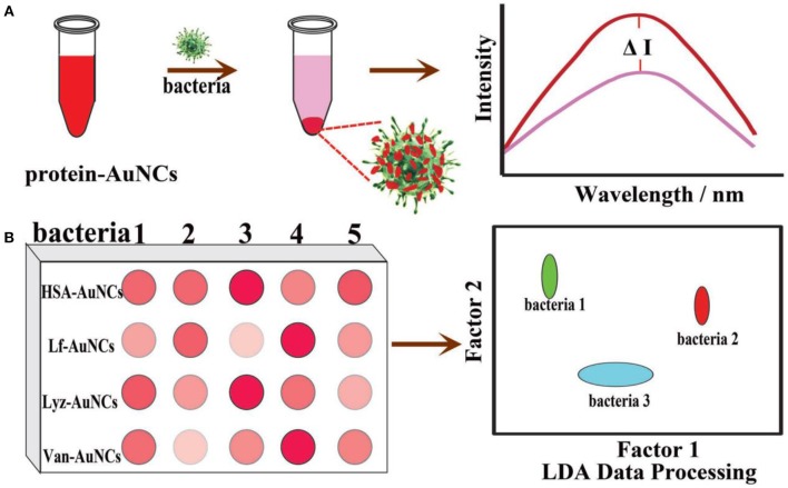
Infections caused by antibiotic-resistant bacteria have become one of the most serious global public health crises. Early detection and effective treatment can effectively prevent deterioration and further spreading of the bacterial infections. Therefore, there is an urgent need for time-saving diagnosis as well as therapeutically potent therapy approaches. Development of nanomedicine has provided more choices for detection and therapy of bacterial infections. Ultrasmall gold nanoclusters (Au NCs) are emerging as potential antibacterial agents and have drawn intense attention in the biomedical fields owing to their excellent biocompatibility and unusual physicochemical properties. Recent significant efforts have shown that these versatile Au NCs also have great application potential in the selective detection of bacteria and infection treatment. In this review, we will provide an overview of research progress on the development of versatile Au NCs for bacterial detection and infection treatment, and the mechanisms of action of designed diagnostic and therapeutic agents will be highlighted. Based on these cases, we have briefly discussed the current issues and perspective of Au NCs for bacterial detection and infection treatment applications.

Keywords: antibacterial activity; bacteria detection; gold nanoclusters; multidrug-resistant bacteria; photoluminescence.


Figures

Scheme 1
Scheme 1
An overview of Au NCs-based bacterial infection diagnostic and therapeutic strategies.
Figure 1
Figure 1
(A) Luminescent HSA-Au NCs as selective probes for Staphylococcus aureus and MRSA. Reproduced from Chan and Chen (2012) with permission from American Chemical Society. (B) Schematic illustration of the working principle for the Cu2+ mediated on-off-on Au NC-based fluorescent probe for rapid Escherichia coli detection. Reproduced from Yan et al. (2018) with permission from American Chemical Society. (C) Simplified scheme of pH controllable adherence of CP-GNC to E. coli cells. Specially, CP-GNC was fully attached to the cells at pH 5.2, whereas all the CP-GNC detached from the surface of E. coli cells at pH 7.4. (D) Bacterial cells can be efficiently labeled and form cell clusters using CP-GNC. Reproduced from Liu P. et al. (2015) with permission from Wiley-VCH Verlag & Co. KGaA, Weinheim.
Figure 2
Figure 2
(A) Schematic illustration of the syntheses of AuAg NCs and photoluminescent quenching by Acinetobacter baumannii. (B) The fluorescence of AuAg NCs was selectively quenched by A. baumannii. The order numbers from 1 to 10 in turn represent the group in the presence of no bacteria (control), Bacillus mycoides, Staphylococcus aureus, methicillin-resistant S. aureus, Candida albicans, P. aeruginosa, E. coli, vancomycin-resistant Enterococcus faecium, Saccharomyces cerevisiae, and A. baumannii, respectively. (C) The concentration dependent quenching effect of A. baumannii toward AuAg NCs. Insets: Digital photos of AuAg NCs under UV illumination after treatment with different concentrations A. baumannii. (D) Relative fluorescence intensity (I0-I) of AuAg NCs in contrast to the logarithm of the A. baumannii concentrations. (E) The transmission electron microscopy (TEM) micrographs of photoluminescent AuAg NCs treated with 1 × 10⁵ CFU/mL A. baumannii demonstrate the bacteria induced agglomeration of NCs. Reproduced from Zheng et al. (2018d) with permission from Elsevier Ltd.
Figure 3
Figure 3
(A) Interaction of the fluorescent probe with bacterial cells: structure of the probe with AHL signal molecules deployed on the surface with lactone and amide moieties intact (top) and specific binding of AHL head groups to receptor sites in Lux-R regulators within bacteria (bottom). Reproduced from Mukherji et al. (2013) with permission from American Chemical Society. (B) Visualization of mannose-protected Au NCs (25 nM) in the absence (left) and presence (right) of E. coli (2.5 × 10⁸ CFU/mL) upon excitation under a hand-held UV lamp (365 nm). Reproduced from Tseng et al. (2011) with permission from Elsevier B.V. (C) Schematic diagram of the synthesis of the red fluorescent lysozyme-Au NCs and fluorescence enhancement detection of E. coli. Reproduced from Liu J. et al. (2015) with permission from Elsevier B.V.
Figure 4
Figure 4
(A) Schematic illustrations of (top) one-step preparation of Au NCs@Van and (bottom) determination of S. aureus in mixtures using the aptamer-coated magnetic beads and Au NCs@Van dual recognition strategy. Reproduced from Cheng et al. (2016) with permission from American Chemical Society. (B) Illustration of the vancomycin and aptamer dual-recognition molecule based FRET assay platform for S. aureus. Reproduced from Yu et al. (2017) with permission from American Chemical Society. (C) Illustration of the immunoassay of E. coli O157:H7 using Au NCs@CS nanocapsules and Au NCs as labels. Reproduced from Cheng et al. (2018) with permission from The Royal Society of Chemistry.
Figure 5
Figure 5
Schematic illustration of protein-Au NC-based fluorescence sensor array for discrimination of various bacteria. (A) The fluorescence intensity of protein-Au NCs was significantly reduced in the presence of bacteria. (B) A schematic fluorescence pattern generated from the different responses of the protein-Au NCs probes toward bacteria. Reproduced from Ji et al. (2018) with permission from Wiley-VCH Verlag & Co. KGaA, Weinheim.
Figure 6
Figure 6
(A) Synthesis of photoluminescent SFT/DT-Au NDs. (B) Comparison of MICs (in terms of the concentration of SFT) of SFT, SFT0.05/DT-Au NDs, SFT0.1/DT-Au NDs, SFT0.25/DT-Au NDs, SFT0.5/DT-Au NDs, and SFT1.0/DT-Au NDs against E. coli, P. vulgaris, MRSA, S. aureus, and Salmonella enterica, respectively. Error bars represent the standard deviation of three repeated measurements. Reproduced from citetbib9 with permission from Wiley-VCH Verlag & Co. KGaA, Weinheim.
Figure 7
Figure 7
(A) Schematic illustrations of the conjugation strategy for antibacterial Au NCs and Dap, conjugation-induced aggregation-induced emission enhancement, and antibacterial synergistic effect. Reproduced from Zheng Y. et al. (2019) with permission from Elsevier Inc. (B) Schematic illustration of antimicrobial peptide-reduced Au NCs with charge-reversal moieties for antibacterial application. Reproduced from Pranantyo et al. (2019) with permission from American Chemical Society.
Figure 8
Figure 8
(A) Schematic illustration of the size regulation of Au NPs to significantly affect their antibacterial properties. Reproduced from Zheng K. et al. (2017) with permission from American Chemical Society. (B) Antibacterial activities of mercaptopyrimidine-conjugated Au NCs indicated with MIC (μg/mL). Here DAMPAu(I) is the precursor complex during the synthesis of Au NCs. Reproduced from Zheng et al. (2018c) with permission from American Chemical Society. (C) Surface ligand chemistry of gold nanoclusters determines their antimicrobial ability. Reproduced from Zheng et al. (2018a) with permission from American Chemical Society.
Figure 9
Figure 9
QA-Au NCs combat bacteria through a multipath mechanism. (A) Scanning electron microscopy (SEM) and (B) TEM images showing the morphological changes of S. aureus after treatment with QA-Au NCs. The administration of QA-Au NCs leads to an increase in the membrane permeability (C), a dissipation of the membrane potential (D) and the generation of ROS (E). The intracellular ATP level (F) and F-type ATPase activity (G) of S. aureus decrease upon treatment with increasing concentrations of QA-Au NCs. Reproduced from Xie et al. (2018) with permission from Wiley-VCH Verlag & Co. KGaA, Weinheim.
Figure 10
Figure 10
(A) Schematic illustration of DPAu/AMD as an image-guided nanotheranostic agent. Reproduced from Setyawati et al. (2014) with permission from American Chemical Society. (B) A simple model representing the possible correlation between the packing density of lipid A of lipopolysaccharide and sepsis progression in the presence of Au NCs. Reproduced from Liao et al. (2018) with permission from American Chemical Society.
Figure 11
Figure 11
(A) Schematic representation of the formation of AuNCs/CS nanoaggregates. (B) The viability of E. coli and S. aureus post treatment with nanoaggregates. Reproduced from Girija et al. (2019) with permission from Wiley-VCH Verlag & Co. KGaA, Weinheim.

References

    1. Alsaiari S. K., Hammami M. A., Croissant J. G., Omar H. W., Neelakanda P., Yapici T., et al. (2017). Colloidal gold nanoclusters spiked silica fillers in mixed matrix coatings: simultaneous detection and inhibition of healthcare-associated infections. Adv. Healthcare Mater. 6:1601135 10.1002/adhm.201601135 - DOI - PubMed
    1. Blair J. M., Webber M. A., Baylay A. J., Ogbolu D. O., Piddock L. J. (2015). Molecular mechanisms of antibiotic resistance. Nat. Rev. Microbiol. 13, 42–51. 10.1038/nrmicro3380 - DOI - PubMed
    1. Boda S. K., Broda J., Schiefer F., Weber-Heynemann J., Hoss M., Simon U., et al. (2015). Cytotoxicity of ultrasmall gold nanoparticles on planktonic and biofilm encapsulated gram-positive staphylococci. Small 11, 3183–3193. 10.1002/smll.201403014 - DOI - PubMed
    1. Boucher H. W., Talbot G. H., Bradley J. S., Edwards J. E., Gilbert D., Rice L. B., et al. (2009). Bad bugs, no drugs: no ESKAPE! An update from the infectious diseases society of America. Clin. Infect. Dis. 48, 1–12. 10.1086/595011 - DOI - PubMed
    1. Chan P. H., Chen Y. C. (2012). Human serum albumin stabilized gold nanoclusters as selective luminescent probes for Staphylococcus aureus and methicillin-resistant Staphylococcus aureus. Anal. Chem. 84, 8952–8956. 10.1021/ac302417k - DOI - PubMed

LinkOut - more resources