. 2021 May;39(8):2679-2692.
doi: 10.1080/07391102.2020.1753577. Epub 2020 Apr 20.

Targeting SARS-CoV-2: a systematic drug repurposing approach to identify promising inhibitors against 3C-like proteinase and 2'-O-ribose methyltransferase

Affiliations

Targeting SARS-CoV-2: a systematic drug repurposing approach to identify promising inhibitors against 3C-like proteinase and 2'-O-ribose methyltransferase

Rameez Jabeer Khan et al. J Biomol Struct Dyn. 2021 May.

Abstract

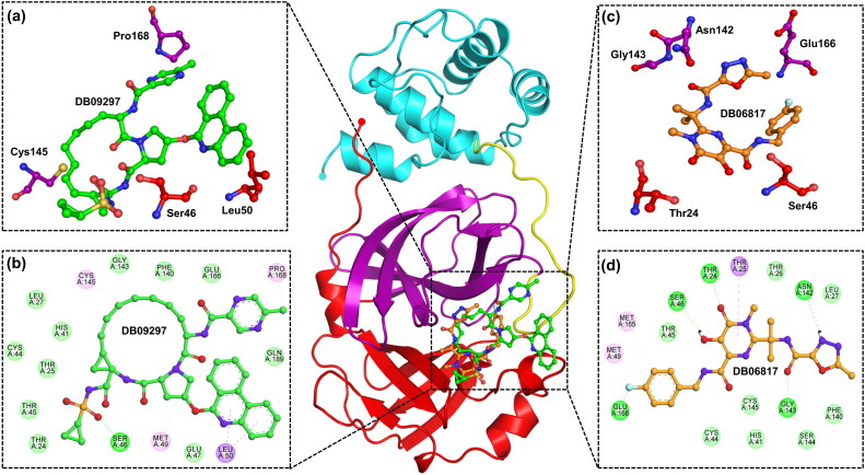
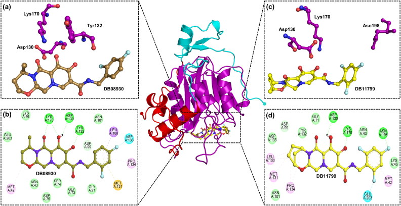
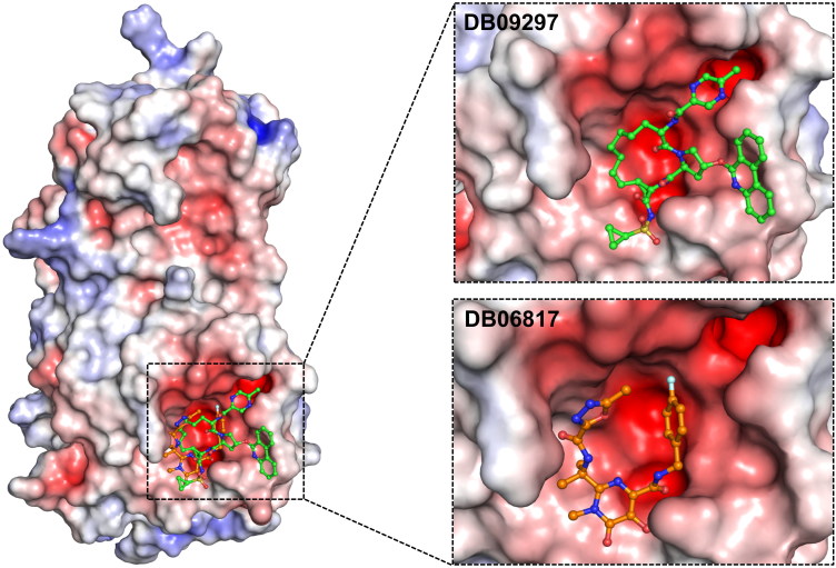
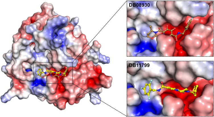
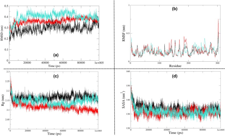
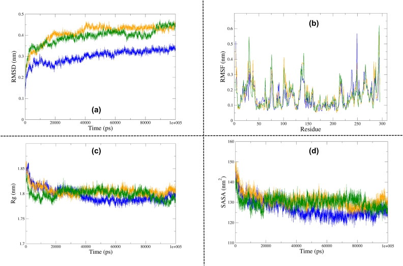
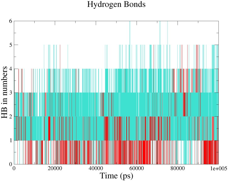
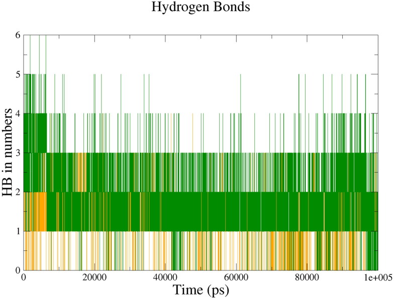
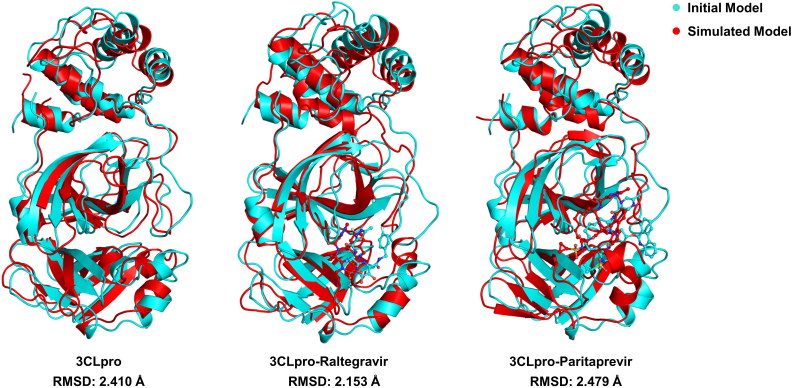
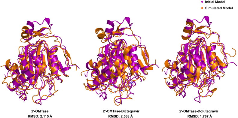
The recent pandemic associated with SARS-CoV-2, a virus of the Coronaviridae family, has resulted in an unprecedented number of infected people. The highly contagious nature of this virus makes it imperative for us to identify promising inhibitors from pre-existing antiviral drugs. Two druggable targets, namely 3C-like proteinase (3CLpro) and 2'-O-ribose methyltransferase (2'-O-MTase) were selected in this study due to their indispensable nature in the viral life cycle. 3CLpro is a cysteine protease responsible for the proteolysis of replicase polyproteins resulting in the formation of various functional proteins, whereas 2'-O-MTase methylates the ribose 2'-O position of the first and second nucleotide of viral mRNA, which sequesters it from the host immune system. The selected drug target proteins were screened against an in-house library of 123 antiviral drugs. Two promising drug molecules were identified for each protein based on their estimated free energy of binding (ΔG), the orientation of drug molecules in the active site and the interacting residues. The selected protein-drug complexes were then subjected to MD simulation, which consists of various structural parameters to equivalently reflect their physiological state. From the virtual screening results, two drug molecules were selected for each drug target protein [Paritaprevir (ΔG = -9.8 kcal/mol) & Raltegravir (ΔG = -7.8 kcal/mol) for 3CLpro and Dolutegravir (ΔG = -9.4 kcal/mol) and Bictegravir (ΔG = -8.4 kcal/mol) for 2'-OMTase]. After the extensive computational analysis, we proposed that Raltegravir, Paritaprevir, Bictegravir and Dolutegravir are excellent lead candidates for these crucial proteins and they could become potential therapeutic drugs against SARS-CoV-2. Communicated by Ramaswamy H. Sarma.

Keywords: 2′-O-ribose methyltransferase; 3C-like proteinase; Drug repurposing; MD simulation; SARS-CoV-2; docking.


Figures

Figure 1.
Figure 1.
Interaction of drugs with 3CLpro (Domain I-Red, Domain II-Purple, Domain III-Cyan, Extended loop-Yellow). (a) Three dimensional representation of 3CLpro active site residues interacting with Paritaprevir (DB09297) (Green). (b) Two dimensional representation of 3CLpro active site residues interacting with Paritaprevir (Green) via Van der Waals interactions (slightly green colour), hydrogen bonds (dark green colour), and pi-interactions (light pink colour). (c) Three dimensional representation of 3CLpro active site residues interacting with Raltegravir (DB06817) (Orange). (d) Two dimensional representation of 3CLpro active site residues interacting with Raltegravir(Orange).
Figure 2.
Figure 2.
Interaction of drugs with 2′-OMTase (MTase Domain-Purple). (a) Three dimensional representation of 2′-OMTase active site residues interacting with Dolutegravir (DB08930) (Sand colour). (b) Two dimensional representation of 2′-OMTase active site residues interacting with Dolutegravir (Sand colour) via Van der Waals interactions (slightly green colour), hydrogen bonds (dark green colour), and pi-interactions (light pink colour, orange colour). (c) Three dimensional representation of 2′-OMTase active site residues interacting with Bictegravir (DB11799) (Yellow). (d) Two dimensional representation of 2′-OMTase active site residues interacting withBictegravir (Yellow).
Figure 3.
Figure 3.
The electrostatic surface potential interaction of Paritaprevir (DB09297) (Green) and Raltegravir (DB06817) (Orange) bound to 3CLpro. The zoomed view is representing the active site cleft.
Figure 4.
Figure 4.
The electrostatic surface potential interaction of Dolutegravir (DB08930) (Sand) and Bictegravir (DB11799) (Yellow) bound to 2′-OMTase. The zoomed view is representing the active site cleft.
Figure 5.
Figure 5.
Analysis of Molecular Dynamics Simulation results of free 3CLpro (Black), 3CLpro-Raltegravir complex (Red) and 3CLpro-Paritaprevir complex (turquoise). (a) Root Mean Square Deviation (RMSD), (b) Root Mean Square Fluctuation (RMSF), (c) Radius of Gyration (Rg), (d) Solvent Accessible Surface Area (SASA).
Figure 6.
Figure 6.
Analysis of Molecular Dynamics Simulation results of free 2′-OMTase (Blue), 2′-OMTase-Bictegravir complex (Orange) and 2′-OMTase-Dolutegravir complex (Dark Green). (a) Root Mean Square Deviation (RMSD), (b) Root Mean Square Fluctuation (RMSF), (c) Radius of Gyration (Rg), (d) Solvent Accessible Surface Area (SASA).
Figure 7.
Figure 7.
Intermolecular hydrogen bonds between the drugs and 3CLpro; 3CLpro-Paritaprevir complex (turquoise), 3CLpro-Raltegravir complex (Red).
Figure 8.
Figure 8.
Intermolecular hydrogen bonds between the drugs and 2′-OMTase; 2′-OMTase-Bictegravir complex (Orange), 2′-OMTase-Dolutegravir complex (Dark Green).
Figure 9.
Figure 9.
Principal Component Analysis (PCA) of free 3CLpro (Black), 3CLpro-Paritaprevir complex (turquoise) and 3CLpro-Raltegravir complex (Red).
Figure 10.
Figure 10.
Principal Component Analysis (PCA) of free 2′-OMTase (Blue), 2′-OMTase-Dolutegravir complex (Dark Green) and 2′-OMTase-Bictegravir complex (Orange).
Figure 11.
Figure 11.
Structural superimposition of the initial and MD simulated free 3CLpro, 3CLpro-Raltegravir complex and 3CLpro-Paritaprevir complex.
Figure 12.
Figure 12.
Structural superimposition of the initial and MD simulated free 2′-OMTase, 2′-OMTase-Bictegravir complex and 2′-OMTase-Dolutegravir complex.

References

    1. Abraham M. J., Lindahl D., van der Spoel E., Hess B., & the GROMACS development team (2014). GROMACS User Manual version, 5(2), 1–298.
    1. Ambure P., Bhat J., Puzyn T., & Roy K. (2019). Identifying natural compounds as multi-target-directed ligands against Alzheimer’s disease: An in silico approach. Journal of Biomolecular Structure and Dynamics, 37(5), 1282–1306. doi:10.1080/07391102.2018.1456975 - DOI - PubMed
    1. Amera G. M., Khan R. J., Pathak A., Jha R. K., Muthukumaran J., & Singh A. K. (2019). Screening of promising molecules against MurG as drug target in multi-drug-resistant-Acinetobacter baumannii – Insights from comparative protein modeling, molecular docking and molecular dynamics simulation. Journal of Biomolecular Structure and Dynamics, 1–23. doi:10.1080/07391102.2019.1700167 - DOI - PubMed
    1. Amera G. M., Khan R. J., Pathak A., Kumar A., & Singh A. K. (2019). Structure based in-silico study on UDP-N-acetylmuramoyl-L-alanyl-D-glutamate-2,6-diaminopimelate ligase (MurE) from Acinetobacter baumannii as a drug target against nosocomial infections. Informatics in Medicine Unlocked, 16, 100216. doi:10.1016/j.imu.2019.100216 - DOI
    1. Benkert P., Biasini M., & Schwede T. (2011). Toward the estimation of the absolute quality of individual protein structure models. Bioinformatics, 27(3), 343–350. doi:10.1093/bioinformatics/btq662 - DOI - PMC - PubMed