. 2020 Jun;128(6):392-402.
doi: 10.1002/cncy.22271. Epub 2020 Apr 8.

Cytologic grading of primary malignant salivary gland tumors: A blinded review by an international panel

Affiliations

Cytologic grading of primary malignant salivary gland tumors: A blinded review by an international panel

Daniel N Johnson et al. Cancer Cytopathol. 2020 Jun.

Abstract

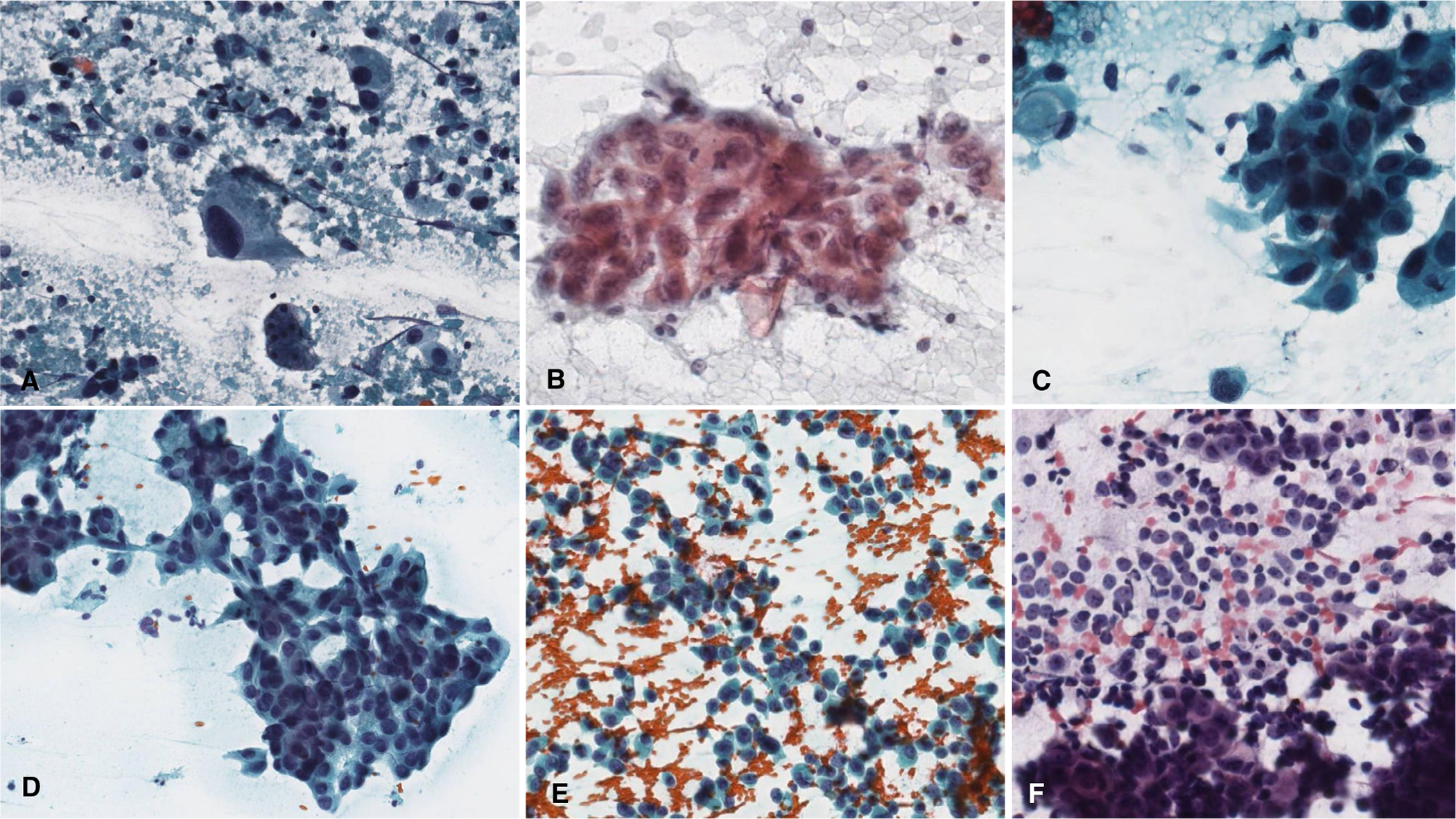
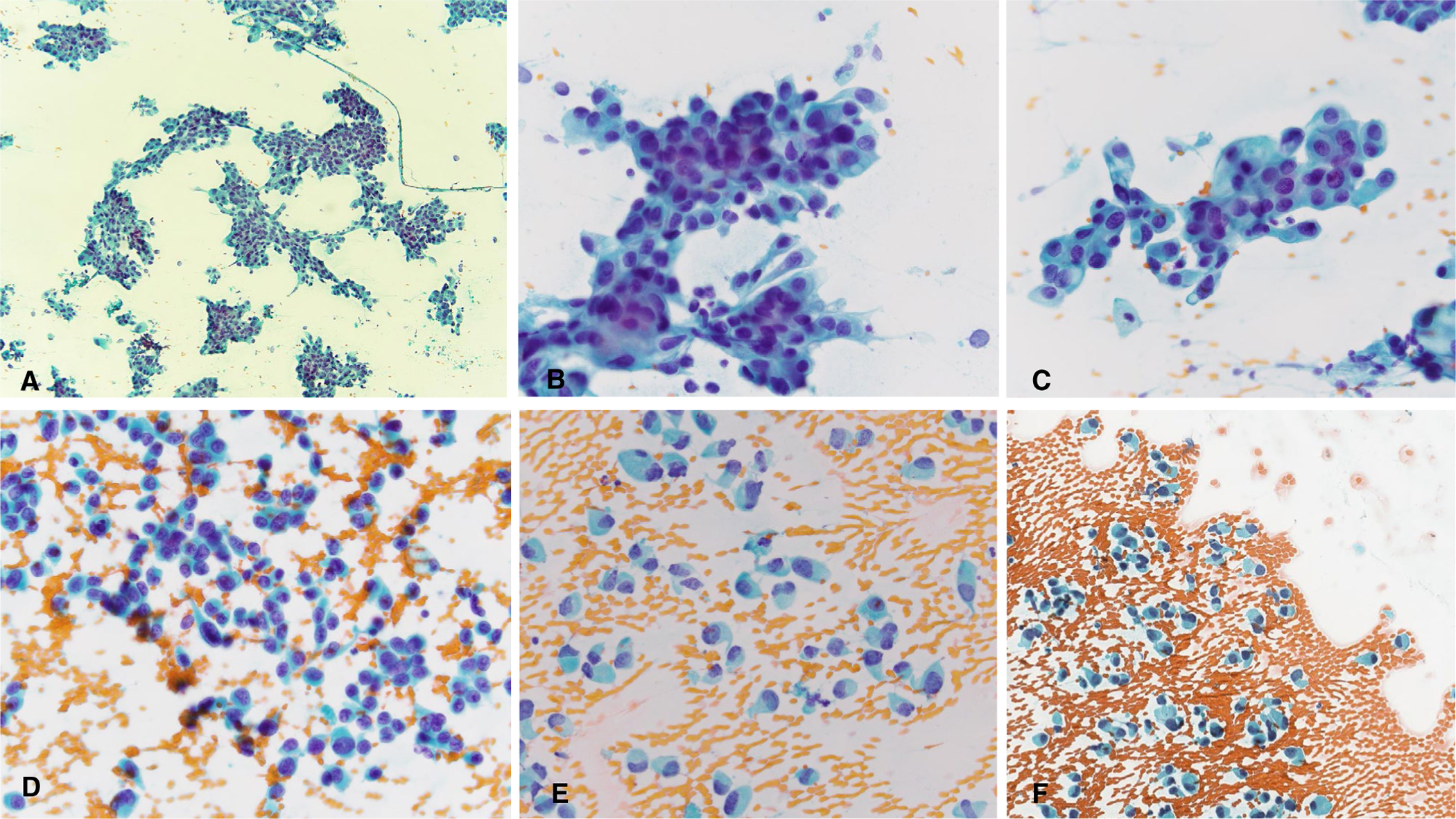
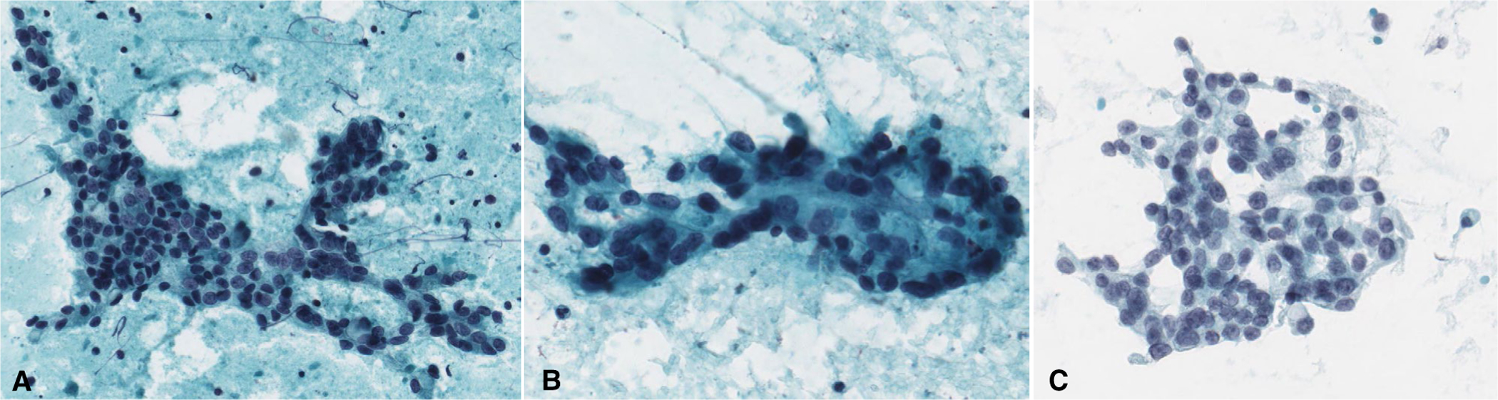
Background: Fine needle aspiration (FNA) is commonly used for the preoperative evaluation of salivary gland tumors. Tumor grade is a key factor influencing clinical management of salivary gland carcinomas (SGCs). To assess the ability to grade nonbasaloid SGCs in FNA specimens, an international panel of cytopathologists convened to review and score SGC cases.

Methods: The study cohort included 61 cases of primary SGC from the pathology archives of 3 tertiary medical centers. Cases from 2005 to 2016 were selected, scanned, and digitized. Nineteen cytopathologists blinded to the histologic diagnosis reviewed the digitized cytology slides and graded them as low, high, or indeterminate. The panelists' results were then compared to the tumor grades based on histopathologic examination of the corresponding resection specimens.

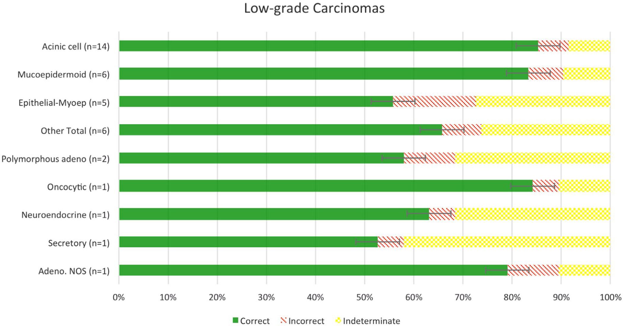
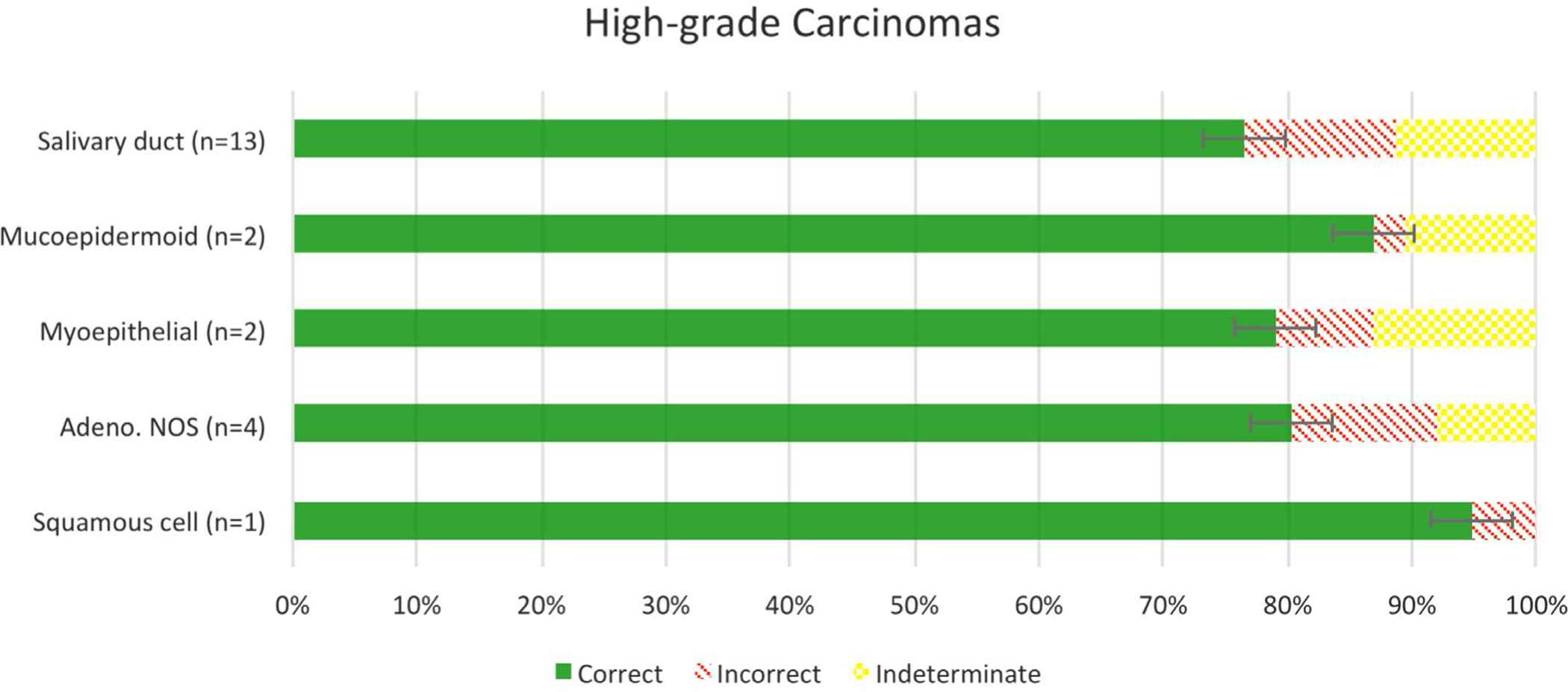
Results: All but 2 of the 19 (89.5%) expert panelists review more than 20 salivary gland FNAs per year; 16 (84.2%) of the panelists work at academic medical centers, and 13 (68.4%) have more than 10 years' experience. Participants had an overall accuracy of 89.4% in the grading of SGC cases, with 90.2% and 88.3% for low- and high-grade SGC, respectively. Acinic cell carcinoma and mucoepidermoid carcinoma had the highest degree of accuracy, while epithelial-myoepithelial carcinoma and salivary duct carcinoma had the lowest degree of accuracy. As expected, the intermediate-grade SGC cases showed the greatest variability (high-grade, 42.1%; low-grade, 37.5%, indeterminate, 20.4%).

Conclusion: This study confirms the high accuracy of cytomorphologic grading of primary SGC by FNA as low- or high-grade. However, caution should be exercised when a grade cannot be confidently assigned.

Keywords: FNA; Milan system; cancer; cytology; grading; salivary gland.


Conflict of interest statement

CONFLICT OF INTEREST DISCLOSURES

The authors made no disclosures.

Figures

FIGURE 1.
FIGURE 1.
Accuracy of grading of low-grade salivary gland carcinomas.
FIGURE 2.
FIGURE 2.
Low-grade salivary gland carcinomas with high (A-C) and low accuracy (D-F). (A) Low-grade mucoepidermoid carcinoma. (B, C) Acinic cell carcinoma. (D) Epithelial-myoepithelial carcinoma with somewhat irregular organization within a cluster of cells. (E) Epithelial-myoepithelial carcinoma with single cells, some with nuclear hyperchromasia. (F) Acinic cell carcinoma exhibiting a large, irregularly shaped cluster. (A, C-F, Papanicolaou stain, original magnification ×400; B, Diff Quik, original magnification ×400.)
FIGURE 3.
FIGURE 3.
Two difficult low-grade salivary gland carcinoma cases. (A-C) Acinic cell carcinoma with hypercellularity with crowded groups, focal fibrous tissue, and variable hyperchromasia that likely led to overgrading. (D-F) Epithelial-myoepithelial carcinoma with hypercellularity, small crowded groups, background debris, and mild hyperchromasia that likely were problematic for accurate grading. (Papanicolaou stain, A, original magnification ×40, B-F, original magnification ×400.)
FIGURE 4.
FIGURE 4.
Accuracy of grading of high-grade salivary gland carcinomas.
FIGURE 5.
FIGURE 5.
High-grade salivary gland carcinomas with high (A-C) and low (D-F) accuracy. (A) Salivary duct carcinoma with overt pleomorphism, single atypical cells, and background necrosis. (B) High-grade mucoepidermoid carcinoma. (C) Salivary duct carcinoma. (D) Salivary duct carcinoma showing somewhat subtle disorganized clusters known as “drunken honey-combing.” (E) Salivary duct carcinoma with abundant single cells and somewhat uniform-appearing nuclear atypia. (F) Adenocarcinoma, not otherwise specified, high-grade with plasmacytoid single cells potentially mistaken for myoepithelial cells. (Papanicolaou stain, original magnification ×400.)
FIGURE 6.
FIGURE 6.
Two difficult high-grade salivary gland carcinomas. (A-C) Salivary duct carcinoma with low nuclear-cytoplasmic ratios and uniform atypia. (D-F) Salivary duct carcinoma with dispersed cell population, plasmacytoid appearance, low nuclear-cytoplasmic ratios, and uniform atypia. (Papanicolau stain, A, original magnification ×40; B-F, Papanicolaou stain, original magnification ×400.)
FIGURE 7.
FIGURE 7.
Intermediate-grade salivary gland carcinomas. (A) Mucoepidermoid carcinoma. (B, C) Adenocarcinoma not otherwise specified. (Papanicolaou stain, original magnification ×400.)

References

    1. Tyagi R, Dey P. Diagnostic problems of salivary gland tumors. Diagn Cytopathol. 2015;43:495–509. - PubMed
    1. Rooper LM. Challenges in minor salivary gland biopsies: a practical approach to problematic histologic patterns. Head and Neck Pathol. 2019;13:476–484. - PMC - PubMed
    1. Seethala RR. An update on grading of salivary gland carcinomas. Head and Neck Pathol. 2009;3:69–77. - PMC - PubMed
    1. Seethala RR. Basaloid/blue salivary gland tumors. Mod Pathol. 2017;30(suppl 1):S84–S95. - PubMed
    1. Skálová A, Gnepp DR, Lewis JS, et al. Newly described entities in salivary gland pathology. Am J Surg Pathol. 2017;41:e33–e47. - PubMed

Publication types