. 2020 May 27;40(22):4418-4431.
doi: 10.1523/JNEUROSCI.2387-19.2020. Epub 2020 Apr 8.

Venlafaxine Stimulates an MMP-9-Dependent Increase in Excitatory/Inhibitory Balance in a Stress Model of Depression

Affiliations

Venlafaxine Stimulates an MMP-9-Dependent Increase in Excitatory/Inhibitory Balance in a Stress Model of Depression

Seham Alaiyed et al. J Neurosci. .

Abstract

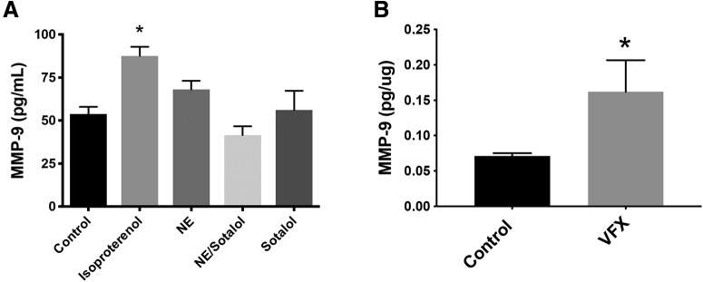
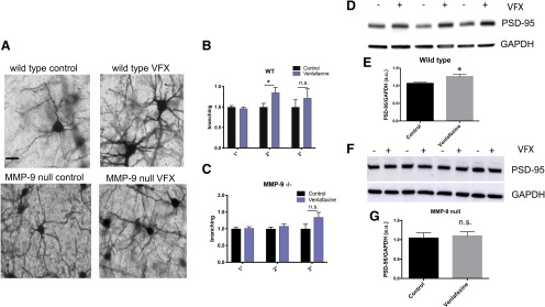
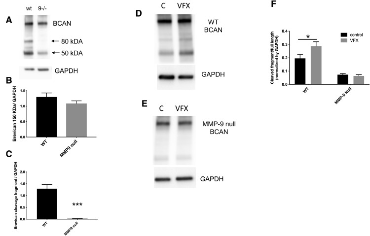
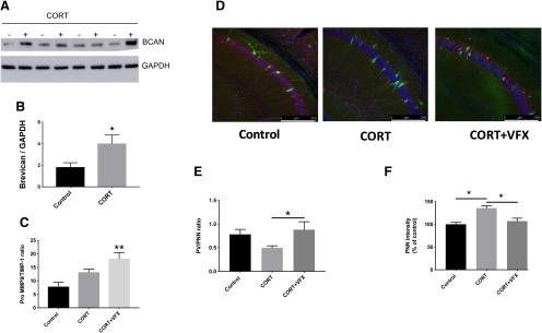
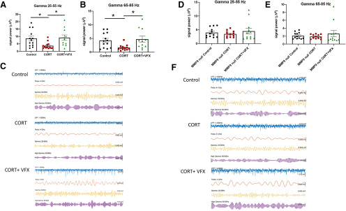
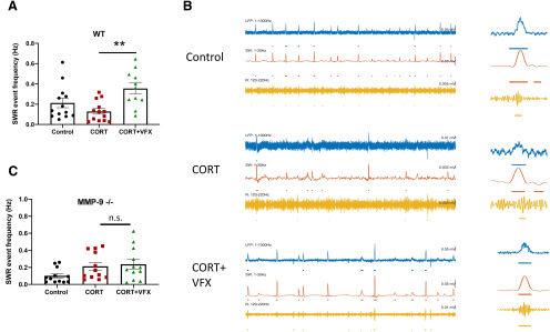
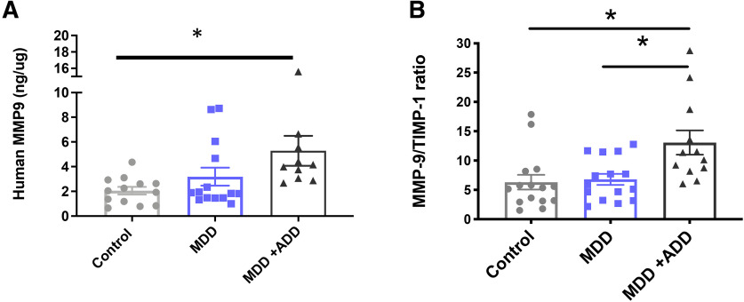
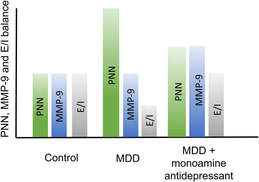
Emerging evidence suggests that there is a reduction in overall cortical excitatory to inhibitory balance in major depressive disorder (MDD), which afflicts ∼14%-20% of individuals. Reduced pyramidal cell arborization occurs with stress and MDD, and may diminish excitatory neurotransmission. Enhanced deposition of perineuronal net (PNN) components also occurs with stress. Since parvalbumin-expressing interneurons are the predominant cell population that is enveloped by PNNs, which enhance their ability to release GABA, excess PNN deposition likely increases pyramidal cell inhibition. In the present study, we investigate the potential for matrix metalloprotease-9 (MMP-9), an endopeptidase secreted in response to neuronal activity, to contribute to the antidepressant efficacy of the serotonin/norepinephrine reuptake inhibitor venlafaxine in male mice. Chronic venlafaxine increases MMP-9 levels in murine cortex, and increases both pyramidal cell arborization and PSD-95 expression in the cortex of WT but not MMP-9-null mice. We have previously shown that venlafaxine reduces PNN deposition and increases the power of ex vivo γ oscillations in conventionally housed mice. γ power is increased with pyramidal cell disinhibition and with remission from MDD. Herein we observe that PNN expression is increased in a corticosterone-induced stress model of disease and reduced by venlafaxine. Compared with mice that receive concurrent venlafaxine, corticosterone-treated mice also display reduced ex vivo γ power and impaired working memory. Autopsy-derived PFC samples show elevated MMP-9 levels in antidepressant-treated MDD patients compared with controls. These preclinical and postmortem findings highlight a link between extracellular matrix regulation and MDD.SIGNIFICANCE STATEMENT Reduced excitatory neurotransmission occurs with major depressive disorder, and may be normalized by antidepressant treatment. Underlying molecular mechanisms are, however, not well understood. Herein we investigate a potential role for an extracellular protease, released from neurons and known to play a role in learning and memory, in antidepressant-associated increases in excitatory transmission. Our data suggest that this protease, matrix metalloprotease-9, increases branching of excitatory neurons and concomitantly attenuates the perineuronal net to potentially reduce inhibitory input to these neurons. Matrix metalloprotease-9 may thus enhance overall excitatory/inhibitory balance and neuronal population dynamics, which are important to mood and memory.

Keywords: MMP; MMP-9; depression; parvalbumin; perineuronal net; venlafaxine.


Figures

Figure 1.
Figure 1.
β-adrenergic receptor activation increases MMP-9 release from cultured neurons, and MMP-9 levels are increased in brain lysates from VFX-treated animals. A, The nonselective β-adrenergic agonist isoproterenol stimulated a significant increase in the release of MMP-9 from cultures of murine hippocampal neurons (n = 5 or 6 biological replicates, p = 0.0013, one-way ANOVA with Tukey's post hoc testing). In addition, while the difference between isoproterenol and norepinephrine was not significant, the difference between isoproterenol and all other groups did reach significance. B, MMP-9 levels in cortical lysates from animals that were treated for 2 weeks with saline or VFX. VFX was associated with a significant increase in cortical MMP-9 (pg/μg total tissue, n = 4 or 5 mice per group, p = 0.044, Student's t test). *p < 0.05.
Figure 2.
Figure 2.
VFX stimulates increased dendritic arbor and PSD-95 levels in WT but not MMP-9-null mice. A, Representative images from Golgi-stained cortex for mouse strains and treatment groups. B, C, Normalized branching data for primary, secondary, and tertiary processes. D-G, Results from PSD-95 quantification in hippocampal lysates. B, C, VFX significantly increases the number of secondary dendrites in WT but not MMP-9-null mice (n = 39-45 neurons from 6-8 mice per group, WT control vs VFX: p = 0.0176, Student's t test; MMP-9-null control vs VFX: not significant). D–G, VFX significantly increased PSD-95 levels in WT mice (p = 0.0388, Student's t test). There was, however, no significant difference in PSD-95 levels between the saline and VFX-treated MMP-9 KOs. Scale bar, 20 μm. *p < 0.05, n.s., non significant.
Figure 3.
Figure 3.
VFX increases BCAN cleavage in WT but not in MMP-9-null mice. A-C, Full-length and cleaved BCAN (80 kDa) in control and MMP-9-null mice. A, Representative Western blot results. B, C, Densitometric results from 4 or 5 mice per group. It can be appreciated that, while full-length BCAN does not significantly differ between WT and MMP-9-null mice (p = 0.24, Student's t test), basal BCAN cleavage is significantly reduced in MMP-9-null animals (p = 0.0004, Student's t test). D-F, Full-length and cleaved BCAN in saline and VFX-treated WT and MMP-9 KO mice. D, E, Representative Western blot results. F, Densitometric analyses with subsequent quantification of the ratio of cleaved/full-length BCAN from n = 4 or 5 mice per group. The difference between control and VFX in WT mice is significant (p = 0.0278, ANOVA with Sidak's multiple comparisons). In contrast, the difference between control and VFX in MMP-9-null animals is not significant (p > 0.05, ANOVA with Sidak's multiple comparisons). *p < 0.05, ***p < 0.001.
Figure 4.
Figure 4.
CORT increases BCAN and PNN levels. A, B, BCAN levels in hippocampal lysates from control and CORT-exposed mice (Western blot and densitometry, respectively). BCAN levels are increased in hippocampal lysates from CORT-exposed mice compared with controls (n = 4 mice per group, p = 0.0466, Student's t test). C, Ratios of MMP-9 and its inhibitor, TIMP-1, in hippocampal lysates from CORT-exposed animals treated with saline or VFX. VFX significantly increased MMP-9/TIMP-1 levels over control (n = 5 or 6 mice per group, p = 0.0027, one-way ANOVA with Tukey's multiple comparisons), whereas CORT alone had no significant effect compared with control (p = 0.094, one-way ANOVA with Tukey's multiple comparisons). D, Representative PNN staining for control, CRT, and CORT + VFX exposed WT animals. E, F, Quantification of the PV/PNN ratio and PNN intensity. VFX significantly increases the PNN/PV ratio in CORT-exposed animals (6-8 mice, n = 18-26 slides, p = 0.0383, ANOVA with Tukey's multiple comparisons) and reduces overall PNN fluorescence intensity (p < 0.0008), consistent with a reduction in PNN levels. Scale bar, 250 μm. *p < 0.05, **p < 0.005.
Figure 5.
Figure 5.
VFX stimulates an MMP-9-dependent increase in the power of γ oscillations in ex vivo hippocampal slices from CORT-treated animals. A, B, Results from LFP recordings of carbachol-stimulated low and high γ power in ex vivo hippocampal slices. VFX increases low and high γ power in CORT-treated animals. The difference between CORT and CORT + VFX groups is significant (4-6 mice, n = 11-13 slices, low γ, p = 0.026, high γ, p = 0.016, one-way ANOVA with Tukey's multiple comparisons), as is the difference between control and CORT groups (low γ, p = 0.041, high γ, p = 0.038, one-way ANOVA with Tukey's multiple comparisons). In contrast, the difference between controls and CORT + VFX groups is not significant. C, Representative tracings from the 3 groups as indicated. D, E, There is no significance difference between the CORT and CORT + VFX groups in MMP-9 KO animals (4 or 5 mice, n = 11 or 12 slices, low γ, p = 0.54, high γ, p = 0.49, one-way ANOVA with Tukey's multiple comparisons). F, We show representative tracings from the KO animals. *p < 0.05.
Figure 6.
Figure 6.
VFX stimulates an MMP-9-dependent increase in SWR abundance in ex vivo hippocampal slices from CORT-treated animals. A, VFX increases SWR abundance in CORT-treated WT animals (4-6 mice, n = 11-13 slices, p = 0.0025, one-way ANOVA with Tukey's multiple comparisons). B, Representative traces for WT animals. C, In contrast, VFX did not significantly increase SWR abundance in CORT-treated MMP-9-null mice (4 or 5 mice, n = 12 slices, p = 0.9, one-way ANOVA with Tukey's multiple comparisons). **p < 0.005, n.s., non significant.
Figure 7.
Figure 7.
Reduced working memory in CORT-treated mice; rescue by VFX in WT mice. VFX reduced closed-arm time in CORT-treated animals tested with the EPM (A; n = 7-10 mice per group, p = 0.029, one-way ANOVA with Sidak's multiple comparisons testing). There was no significant difference between the CORT and CORT + VFX groups in MMP-9-null animals (B; n = 7-10 animals per group, p = 0.84, ANOVA with Tukey's multiple comparisons testing). C, We examined the effects of CORT on T-maze performance. CORT decreased working memory in WT mice (n = 10 mice per group, p = 0.0049, Student's t test). C, D, We also examined working memory in WT and MMP-9-null mice treated with CORT or CORT + VFX. VFX improved T-maze performance in CORT-treated control (n = 5 mice per group, p = 0.045, ANOVA with Sidak's multiple comparisons testing). In addition, VFX improved performance in animals that did not receive CORT (n = 5 mice per group, p = 0.045, ANOVA with Sidak's multiple comparisons testing). The difference between control and CORT groups was not significant in this cohort, which may be due to injection stress in both groups and/or a smaller n. In addition, although the WT controls in C and D showed differential performance, it should be noted that controls in D received stressful daily intraperitoneal saline, whereas controls in C received CD in their drinking water. E, T-maze results for MMP-9-null animals. We did not have a VFX only group for these animals. However, in contrast to WT animals, VFX did not improve performance in CORT-treated MMP-9-null mice (n = 7-10 animals per group, p = 0.36, ANOVA with Tukey's multiple comparisons testing). *p < 0.05, n.s., non significant.
Figure 8.
Figure 8.
MMP-9/TIMP-1 levels are increased in PFC samples from MDD patients treated with monoamine antidepressants. A, B, Compared with control patients, monoamine antidepressant-treated patients (ADD) have increased levels of MMP-9 in the PFC (A, p = 0.0130, one-way ANOVA with Dunnett's multiple comparisons). In contrast, there is no significant difference between MMP-9 levels in control and untreated MDD patients (p = 0.45). Moreover, there is a significant increase in the MMP-9/TIMP-1 ratio in monoamine antidepressant-treated MDD patients compared with the same ratio in controls or untreated MDD patients (Control vs MDD + ADD, p = 0.0052, one-way ANOVA with Tukey's multiple comparisons; MDD vs MDD + ADD, p = 0.009, one-way ANOVA with Tukey's multiple comparisons). *p < 0.05.
Figure 9.
Figure 9.
Hypothetical changes in PNN deposition and MMP-9 activity in untreated and treated MDD. Hypothetical PNN, MMP levels, and E/I balance (arbitrary units) are shown for nondepressed control patients. With MDD, PNN levels increase and thus E/I balance decreases. With MDD in the setting of antidepressant therapy, however, MMP levels also increase to potentially restore E/I balance.

References

    1. Alaiyed S, Bozzelli PL, Caccavano A, Wu JY, Conant K (2019) Venlafaxine stimulates PNN proteolysis and MMP-9-dependent enhancement of gamma power: relevance to antidepressant efficacy. J Neurochem 148:810–821. 10.1111/jnc.14671 - DOI - PMC - PubMed
    1. Alekseichuk I, Turi Z, Amador de Lara G, Antal A, Paulus W (2016) Spatial working memory in humans depends on theta and high gamma synchronization in the prefrontal cortex. Curr Biol 26:1513–1521. 10.1016/j.cub.2016.04.035 - DOI - PubMed
    1. Allen M, Ghosh S, Ahern GP, Villapol S, Maguire-Zeiss KA, Conant K (2016) Protease induced plasticity: matrix metalloproteinase-1 promotes neurostructural changes through activation of protease activated receptor 1. Sci Rep 6:35497 10.1038/srep35497 - DOI - PMC - PubMed
    1. Arikan MK, Metin B, Tarhan N (2018) EEG gamma synchronization is associated with response to paroxetine treatment. J Affect Disord 235:114–116. 10.1016/j.jad.2018.04.041 - DOI - PubMed
    1. Balmer TS. (2016) Perineuronal nets enhance the excitability of fast-spiking neurons. eNeuro 3:ENEURO.0112-16.2016 10.1523/ENEURO.0112-16.2016 - DOI - PMC - PubMed

Publication types

MeSH terms

Substances