. 2020 Jun 1;94(12):e00132-20.
doi: 10.1128/JVI.00132-20. Print 2020 Jun 1.

Swine ANP32A Supports Avian Influenza Virus Polymerase

Affiliations

Swine ANP32A Supports Avian Influenza Virus Polymerase

Thomas P Peacock et al. J Virol. .

Abstract

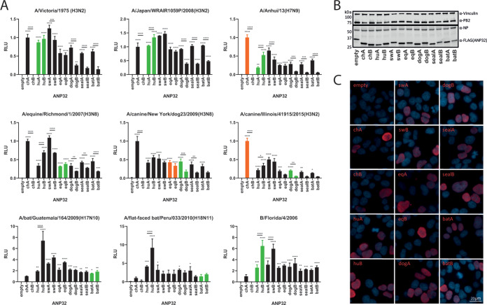
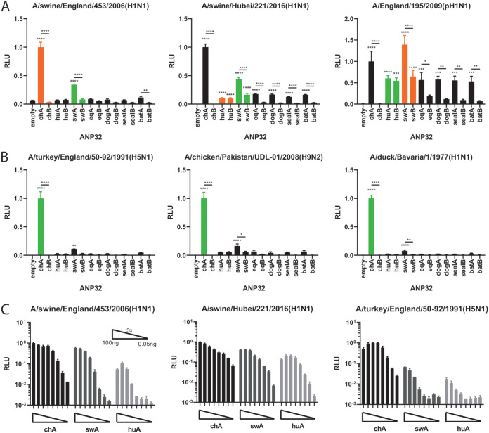
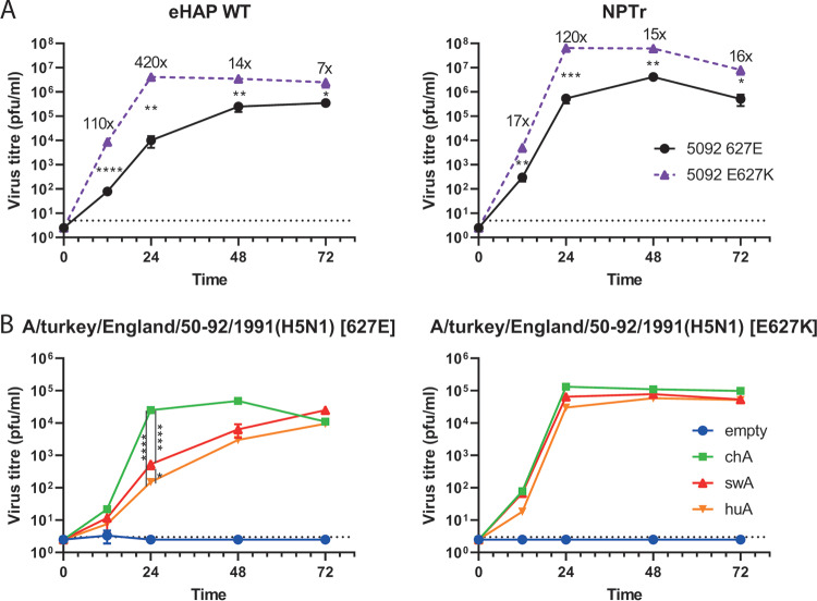
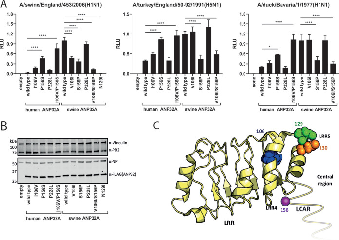
Avian influenza viruses occasionally infect and adapt to mammals, including humans. Swine are often described as "mixing vessels," being susceptible to both avian- and human-origin viruses, which allows the emergence of novel reassortants, such as the precursor to the 2009 H1N1 pandemic. ANP32 proteins are host factors that act as influenza virus polymerase cofactors. In this study, we describe how swine ANP32A, uniquely among the mammalian ANP32 proteins tested, supports the activity of avian-origin influenza virus polymerases and avian influenza virus replication. We further show that after the swine-origin influenza virus emerged in humans and caused the 2009 pandemic, it evolved polymerase gene mutations that enabled it to more efficiently use human ANP32 proteins. We map the enhanced proviral activity of swine ANP32A to a pair of amino acids, 106 and 156, in the leucine-rich repeat and central domains and show these mutations enhance binding to influenza virus trimeric polymerase. These findings help elucidate the molecular basis for the mixing vessel trait of swine and further our understanding of the evolution and ecology of viruses in this host.IMPORTANCE Avian influenza viruses can jump from wild birds and poultry into mammalian species such as humans or swine, but they only continue to transmit if they accumulate mammalian adapting mutations. Pigs appear uniquely susceptible to both avian and human strains of influenza and are often described as virus "mixing vessels." In this study, we describe how a host factor responsible for regulating virus replication, ANP32A, is different between swine and humans. Swine ANP32A allows a greater range of influenza viruses, specifically those from birds, to replicate. It does this by binding the virus polymerase more tightly than the human version of the protein. This work helps to explain the unique properties of swine as mixing vessels.

Keywords: ANP32; ANP32A; ANP32B; host factors; influenza; pandemic; replication; swine; swine influenza; zoonotic.


Figures

FIG 1
FIG 1
Most common mammalian influenza hosts have two ANP32 proteins capable of supporting influenza polymerase. (A) Minigenome assays performed in human eHAP dKO cells with ANP32 proteins from different avian or mammalian species cotransfected. Green bars indicate species from which the influenza virus polymerase was isolated; orange bars indicate recent species from which the virus has jumped. Data indicate triplicate repeats plotted as mean with standard deviation. Data for each polymerase normalized to chicken ANP32A. (B) Western blot assay showing protein expression levels of FLAG-tagged ANP32 proteins, NP, and PB2 during a minigenome assay. (C) Immunofluorescence images showing nuclear localization of all FLAG-tagged ANP32 proteins (red) tested. Nuclei are stained with DAPI (blue). ch, chicken; hu, human; sw, swine; eq, equine. Statistical significance was determined by one-way analysis of variance (ANOVA) with multiple comparisons against empty vector or between ANP32 proteins from the same host. *, 0.05 ≥ P > 0.01; **, 0.01 ≥ P > 0.001; ***, 0.001 ≥ P > 0.0001; ****, P ≤ 0.0001.
FIG 2
FIG 2
swANP32A can support the activity of minimally mammalian-adapted or completely nonadapted polymerases. Minigenome assays of swine (A) and avian (B) polymerases performed in human eHAP dKO cells with ANP32 proteins from different avian or mammalian species cotransfected. Green bars indicate species from which the influenza virus polymerase was isolated; orange bars indicate recent species from which the virus has jumped. Data indicate triplicate repeats plotted as mean with standard deviation. Data for each polymerase normalized to chicken ANP32A. (C) ANP32 protein titrations with three different virus polymerase constellations. ANP32 expression plasmids were diluted in a series of 3× dilutions starting with 100 ng. Data indicate triplicate repeats plotted as mean with standard deviation. Statistical significance was determined by one-way analysis of variance (ANOVA) with multiple comparisons against empty vector. **, 0.01 ≥ P > 0.001; ***, 0.001 ≥ P > 0.0001; ****, P ≤ 0.0001.
FIG 3
FIG 3
Swine ANP32A can support avian influenza virus replication better than human ANP32A. Comparative growth kinetics of isogenic, recombinant avian influenza viruses (A/turkey/England/50-92/1991 [H5N1]) PB2 627E (wild type) versus E627K in wild-type human eHAP cells and swine NPTr cells (A) and eHAP dKO cells (B) preexpressing empty vector, chicken, swine, or human ANP32A. Cells were infected at a multiplicity of infection (MOI) of 0.001. All time points taken in triplicate, and mean viral titers were determined by plaque assay in MDCK cells with the standard deviation shown. Graph shows representative data of at least two independent repeats showing the same trends. Statistical significance determined by multiple Student’s t tests in panel A and one-way analysis of variance (ANOVA) with multiple comparisons in panel B. Value shown on graph in panel A indicate fold change in mean titers. Dotted lines on graphs indicate limits of detection. *, 0.05 ≥ P > 0.01; **, 0.01 ≥ P > 0.001; ***, 0.001 ≥ P > 0.0001; ****, P ≤ 0.0001.
FIG 4
FIG 4
Third-wave pandemic H1N1 viruses adapt to human ANP32 proteins through the PA mutation N321K. (A) Minigenome assays of polymerases derived from first- and third-wave pH1N1 viruses (E195 and E687, respectively) performed in wild-type human eHAP cells and swine NPTr cells. Data indicate triplicate repeats plotted as mean with standard deviation. Data normalized to wild-type pH1N1 E195. (B) Minigenome assays performed in human eHAP cells with ANP32A and ANP32B knocked out and complemented with ANP32 proteins from human or swine following cotransfection of expression plasmids. Data indicate triplicate repeats plotted as mean with standard deviation. Data normalized to pH1N1 E195 wild type with chicken ANP32A. All experiments in panels A and B performed on two separate occasions with a representative repeat shown. (C) Indirect immunofluorescence images showing endogenous nuclear localization of swine ANP32A in swine NPTr cells. Statistical significance was determined by one-way analysis of variance (ANOVA) with multiple comparisons. ****, P ≤ 0.0001.
FIG 5
FIG 5
The enhanced proviral activity of swine ANP32A maps to amino acids in LRR4 and the central domain. (A) Minigenome assays with polymerase constellations from a swine or an avian influenza virus performed in human eHAP dKO cells with human/swine ANP32A reciprocal mutants expressed. Data indicate triplicate repeats plotted as mean with standard deviation repeated on two separate occasions with a representative repeat shown. Data normalized to each polymerase with swine wild-type ANP32A. (B) Western blot analysis showing expression levels of human/swine ANP32A from minigenome assays. (C) Crystal structure of ANP32A (PDB accession no. 2JE1) with residues found to affect proviral activity mapped (39). The unresolved, unstructured LCAR is shown as a yellow line. Schematic made using PyMol (40). Statistical significance was determined by one-way analysis of variance (ANOVA) with multiple comparisons. *, 0.05 ≥ P > 0.01; ***, 0.001 ≥ P > 0.0001; ****, P ≤ 0.0001.
FIG 6
FIG 6
Amino acid residues responsible for the enhanced support of polymerase activity of swine ANP32A also mediate increased binding to influenza trimeric polymerase. (A) Split-luciferase assays showing the relative binding of different ANP32 proteins to trimeric polymerase from human pH1N1 or avian H5N1 viruses. PB1 was tagged with the N-terminal part of Gaussia luciferase, while ANP32 proteins were tagged with the C-terminal part. NLR, normalized luminescence ratio, calculated from the ratio between tagged and untagged ANP32/PB1 pairs. Assay performed in 293T cells. Data indicate triplicate repeats plotted as mean with standard deviation, repeated across two separate experiments with representative data shown. Statistical significance was determined by one-way ANOVA with multiple comparisons between the swA and huA wild types and mutants. ***, 0.001 ≥ P > 0.0001; ****, P ≤ 0.0001. (B) Minigenome assays with reconstituted polymerases from 3 different influenza viruses, performed in human eHAP cells with ANP32A and ANP32B knocked out and complemented with wild-type swine ANP32A or B or N129I mutants thereof. Data indicate triplicate repeats plotted as mean with standard deviation, repeated across two separate experiments with representative data shown. Data normalized to each polymerase with wild-type swine ANP32A. ****, P ≤ 0.0001. (C) Western blot assay showing protein expression levels of FLAG-tagged swine ANP32 wild type or N129I proteins during a minigenome assay. (D) Phylogenetic tree of mammalian ANP32A proteins. Species that contain the highly proviral 156S shown in red; species with 156P shown in black. Phylogenetic trees made using the neighbor-joining method based on amino acid sequence. Statistical significance was determined by one-way analysis of variance (ANOVA) with multiple comparisons against an empty vector.

References

    1. Li S, Shi Z, Jiao P, Zhang G, Zhong Z, Tian W, Long LP, Cai Z, Zhu X, Liao M, Wan XF. 2010. Avian-origin H3N2 canine influenza A viruses in Southern China. Infect Genet Evol 10:1286–1288. doi:10.1016/j.meegid.2010.08.010. - DOI - PMC - PubMed
    1. Kandeil A, Gomaa MR, Shehata MM, El Taweel AN, Mahmoud SH, Bagato O, Moatasim Y, Kutkat O, Kayed AS, Dawson P, Qiu X, Bahl J, Webby RJ, Karesh WB, Kayali G, Ali MA. 2018. Isolation and Characterization of a Distinct Influenza A Virus from Egyptian Bats. J Virol 93:e01059-18. doi:10.1128/JVI.01059-18. - DOI - PMC - PubMed
    1. Geraci JR, St Aubin DJ, Barker IK, Webster RG, Hinshaw VS, Bean WJ, Ruhnke HL, Prescott JH, Early G, Baker AS, Madoff S, Schooley RT. 1982. Mass mortality of harbor seals: pneumonia associated with influenza A virus. Science 215:1129–1131. doi:10.1126/science.7063847. - DOI - PubMed
    1. Taubenberger JK, Reid AH, Lourens RM, Wang R, Jin G, Fanning TG. 2005. Characterization of the 1918 influenza virus polymerase genes. Nature 437:889–893. doi:10.1038/nature04230. - DOI - PubMed
    1. Pensaert M, Ottis K, Vandeputte J, Kaplan MM, Bachmann PA. 1981. Evidence for the natural transmission of influenza A virus from wild ducts to swine and its potential importance for man. Bull World Health Organ 59:75–78. - PMC - PubMed

Publication types

MeSH terms