. 2020 Apr;580(7802):257-262.
doi: 10.1038/s41586-020-2134-y. Epub 2020 Mar 25.

A conserved dendritic-cell regulatory program limits antitumour immunity

Affiliations

A conserved dendritic-cell regulatory program limits antitumour immunity

Barbara Maier et al. Nature. 2020 Apr.

Erratum in

Abstract

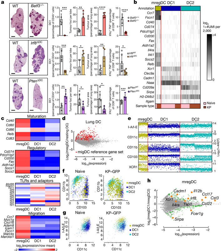
Checkpoint blockade therapies have improved cancer treatment, but such immunotherapy regimens fail in a large subset of patients. Conventional type 1 dendritic cells (DC1s) control the response to checkpoint blockade in preclinical models and are associated with better overall survival in patients with cancer, reflecting the specialized ability of these cells to prime the responses of CD8+ T cells1-3. Paradoxically, however, DC1s can be found in tumours that resist checkpoint blockade, suggesting that the functions of these cells may be altered in some lesions. Here, using single-cell RNA sequencing in human and mouse non-small-cell lung cancers, we identify a cluster of dendritic cells (DCs) that we name 'mature DCs enriched in immunoregulatory molecules' (mregDCs), owing to their coexpression of immunoregulatory genes (Cd274, Pdcd1lg2 and Cd200) and maturation genes (Cd40, Ccr7 and Il12b). We find that the mregDC program is expressed by canonical DC1s and DC2s upon uptake of tumour antigens. We further find that upregulation of the programmed death ligand 1 protein-a key checkpoint molecule-in mregDCs is induced by the receptor tyrosine kinase AXL, while upregulation of interleukin (IL)-12 depends strictly on interferon-γ and is controlled negatively by IL-4 signalling. Blocking IL-4 enhances IL-12 production by tumour-antigen-bearing mregDC1s, expands the pool of tumour-infiltrating effector T cells and reduces tumour burden. We have therefore uncovered a regulatory module associated with tumour-antigen uptake that reduces DC1 functionality in human and mouse cancers.


Conflict of interest statement

Competing interests Research support for these studies was provided by Regeneron and Takeda. The authors declare no other competing financial interests.

Figures

Extended Data Fig. 1 |
Extended Data Fig. 1 |. mregDCs are a distinct dendritic-cell cluster present in numerous tumour models
a, Digested lungs of naive or KP-tumour-bearing mice at day 28 post tumour-cell injection were stained with antibodies conjugated either to fluorophores for FACS or to oligonucleotides for CITE-seq analysis. CD45+ Siglec F Ly6G MHCII+ CD11c+ cells were sorted and loaded onto a 10x Chromium chip for scRNA-seq and CITE-seq analysis. Dendritic-cell clusters were identified according to marker-gene expression after clustering of transcriptomes. Heat maps show UMI counts of lineage genes across all clusters after downsampling to 2,000 UMIs per cell. b, Left, gene–gene correlation of highly variable genes, with relevant gene modules outlined and annotated; right, scRNA expression divided by cluster. Genes on left and right panels are aligned. c, CD45+ Siglec F Ly6G MHCII+ CD11c+ cells from lungs of naive or B16-BFP/OVA-tumour-bearing mice at day 22 were sorted and loaded onto a 10x Chromium platform for scRNA-seq. DCs were mapped to the clusters generated for the experiment shown inFig. 1 by maximum-likelihood classification. Heat maps show UMI counts of lineage genes across all clusters after downsampling to 2,000 UMIs per cell. d, Mouse-tumour public scRNA data for immune cells from an M38 model and a T3 sarcoma model were accessed from GEO. Top, broad cell types were sorted in silico using gene lists, resulting in pure DC populations. pDC, plasmacytoid DC. Bottom left, DC1s, DC2s and mregDCs were identified using scores generated from gene lists that defined these populations. Bottom middle, annotations in the heat map were derived from k-means clustering (k = 3) of coordinates in the dendritic-cell-score scatter plot. Bottom right, DCs of each annotation are quantified. Gene lists defining cell types for in silico sorting and stratification of dendritic-cell subtypes are in Supplementary Table 2. e, Lung DC1s and migratory DC1s (migDC1) from DLNs were sorted and analysed by RNA-seq. Genes highlighted in red identify a reference set of genes from migratory DCs. f, Lung DC1s and migratory DC1s from DLNs in both naive and KP-tumour-bearing mice were sorted and analysed by RNA-seq. The plot compares migDC1 gene expression with lung DC1 expression by log2FC in naive (x-axis) and KP-tumour-bearing (y-axis) mice. Genes upregulated in mregDCs relative to DC1s (log2FC greater than 2; Benjamini–Hochberg-adjusted P-value less than 0.01), as assayed by scRNA-seq, are shown in gold. g, Stratification of dendritic-cell transcriptomes using dendritic-cell subtype scores in naive and KP-tumour-bearing lungs. Scores for each subtype were generated from gene lists that were differentially expressed among clusters. Single cells are coloured by cluster identification (left) or CITE-seq surface marker expression (colour-bar units are log10(1 + ADT counts)). Gene scores are the same as in d (lower left).
Extended Data Fig. 2 |
Extended Data Fig. 2 |. The mregDC program is enriched in both canonical dendritic-cell subsets upon tumour-antigen uptake.
a, b, CD45+ Siglec F Ly6G MHCII+ CD11c+ cells were sorted from lungs of Ccr7−/− mice and loaded onto the 10x Chromium followed by scRNA-seq. Transcriptomes were mapped to the clusters generated for the wild-type experiment shown in Fig. 1 by maximum-likelihood classification. a, The heat map shows UMI counts of selected genes in dendritic-cell clusters after downsampling to 2,000 UMIs per cell, comparing cells from Ccr7−/− mice to cells from WT mice. b, Comparison of differential expression analyses between mregDCs and resting DCs in WT mice (x-axis) and Ccr7−/− mice ( y-axis) (b). c, Frequencies of mregDCs as a percentage of total DCs, as measured by scRNA-seq in naive and KP–GFP-tumour-bearing mice. d, Gating strategy for subsets of conventional lung DCs. e, Flow cytometry of GFP+ versus GFP DC2s (CD11b+ CD103) from KP–GFP-tumour- bearing mice. f, Flow cytometry of GFP+ versus GFP- DC1s or DC2s from KP–GFP-tumour-bearing mice. g, Flow cytometry of BFP+ versus BFP DC1s from B16-BFP/OVA tumour-bearing mice. The experiment shown is representative of two independent experiments; *P < 0.05; **P < 0.01, ***P < 0.001, ****P < 0.0001 (Student’s t-test); data are means ± s.d. (e–g). h, KP–GFP cells were exposed to ultraviolet radiation for 30 min, rested for 24 h, and stained with annexin V and propidium iodide in order to confirm induction of apoptosis before experiments involving coculture of DCs. i, Differential expression between mregDCs identified by transcriptome from KP-tumour-bearing and naive mice. Genes in green are significantly differentially expressed (Benjamini–Hochberg-adjusted P-value of less than 0.15); selected immune genes are shown in orange.
Extended Data Fig. 3 |
Extended Data Fig. 3 |. The mregDC program is independent of MyD88/TRIF, inflammasome signalling and lymphocytes
a–d, Flow-cytometry analysis of DC1s isolated from KP–GFP-tumour-bearing lungs in Asc−/− (a), Il1r−/− (b), Myd88−/−Trif−/− (c) and Rag1−/− (d) mice. e, Heat map showing average TAM receptor RNA expression in mregDC, DC1 and DC2 scRNA-seq clusters. f, Differential expression of TH2 response genes across dendritic-cell clusters identified by scRNA-seq, showing a log2FC between average mregDC expression and resting dendritic-cell expression. Genes in green are differentially expressed (Benjamini–Hochberg-adjusted P-value less than 0.15); TH2 response genes are in orange. g, k, Mice were injected with KP–GFP tumour cells, treated with anti-IL-4 (αIL-4) or control IgG on days 21, 24 and 26, and analysed on day 28. GFP+ DC1s carrying tumour antigens in lung and DLNs (g) and T cells in DLNs (k) were analysed by flow cytometry. h, KP–GFP-tumour-bearing mice were injected with an agonistic CD40 antibody (CD40a) on days 25 and 27; lungs were analysed on day 28. i, KP–GFP-tumour-bearing mice were injected with polyI:C on day 27, and lungs were analysed on day 28.j, GFP+ conventional DC1s were purified from KP–GFP-tumour-bearing lungs from B6D2 mice treated either with anti-IL-4 or control IgG and cocultured with naive CD8+ JEDI T cells isolated from JEDI mouse spleens. JEDI T cells were analysed on day 2. One experiment, representative of two independent experiments, is shown (a–d, g–k). *P < 0.05; **P < 0.01; ***P < 0.001; ****P < 0.0001 (Student’s t-test (a–d, g, k) or one-way ANOVA and Tukey’s test (h, i)). Data are shown as means ± s.d. (a–d, g–i, k).
Extended Data Fig. 4 |
Extended Data Fig. 4 |. Protein expression profile of mregDCs in human NSCLC lesions
a, Average CITE-seq surface protein staining intensity of dendritic-cell clusters in non-involved lung (nLung) and tumour lesions isolated from human NSCLC resections (n = 7). b, scRNA-seq data from a published dataset of matched nLung and tumour from resection specimens of eight patients with NSCLC were mapped to the clusters generated for the NSCLC data in Fig. 4 by maximum-likelihood classification. Heat maps show downsampled UMI counts in dendritic-cell clusters after downsampling cells to 2,000 UMIs per cell and evenly sampling cells from dendritic-cell types.
Fig. 1 |
Fig. 1 |. Identification of a dendritic-cell cluster enriched in immunoregulatory and maturation molecules.
a, Lung tumours were quantified in Batf3+/+ or Batf3−/−, Irf8WT or Irf8δDC, and PtenWT or PtenδDC mice (DC, dendritic cell; WT, wild type). At the left are images of the right-hand lungs; scale bar, 1 mm. At the right are quantifications (by flow cytometry) of DC1s as a proportion of CD45+ (immune) cells; tumour foci and area; and IFNγ+ TNF+ CD8+ T cells. Right lungs were digested for flow cytometry. The results shown are from one experiment, representative of three independent experiments (with five mice per experiment). *P < 0.05; **P < 0.01; ***P < 0.001; ****P < 0.0001 (Student’s t-test). Data are shown as means ± standard deviation (s.d.). bh, CD45+ lin MHCII+ CD11c+ cells from lungs of naive or KP-tumour-bearing mice were sorted for scRNA-seq and CITE-seq. b, c, Heat maps show unique molecular identifier (UMI) counts of selected genes, with key indicating sample type of origin (b), or relative cluster averages (c). d, Differential expression between mregDC and average tissue-resident dendritic cell (pooled DC1 and DC2) signatures, with genes belonging to a reference migratory dendritic-cell signature indicated in red. e–g, Protein expression levels detected by CITE-seq, grouped by transcriptome-defined cluster. h, Differential gene expression between mregDC1s versus mregDC2s in naive lungs. Genes indicated in green are significant with Benjamini–Hochberg-adjusted P-values of less than 0.15.
Fig. 2 |
Fig. 2 |. The mregDC1 program is associated with uptake of tumour antigens.
a, b, CD45+ lin MHCII+ CD11c+ cells were sorted from the lungs of Ccr7−/− mice for scRNA-seq. Expression profiles (a) and frequencies of mregDCs as a proportion of total dendritic cells (b) in WT and Ccr7−/− mice are shown. c, Flow cytometry of lungs and DLNs from WT mice bearing KP–GFP tumours. Results shown are from one experiment, representative of three independent experiments (n = 5). d, CD45+ lin MHCIIhi CD11c+ CD24hi CD11b CD103+ cells from WT mouse lungs were sorted using fluorescence-activated cell sorting and stained for EEA1 (an endosomal marker). e, Lung GFP+ and GFP DC1 populations were sorted from mice bearing KP–GFP tumours and analysed by RNA-seq. Genes that are upregulated in mregDCs relative to DC1s (with a log2-transformed fold change (log2FC) of more than 2; Benjamini–Hochberg-adjusted P-value of less than 0.01) are shown in gold. P-values of signature association are less than 2.2 × 10⁻¹⁶ (Fisher’s exact test). f, Flow cytometry of DC1s from mouse lungs bearing KP–GFP tumours. g, Ultraviolet-irradiated KP–GFP cells were added to a bone-marrow-derived DC1 culture for 2 h before analysis of DC1s by flow cytometry. h, GFP+ and GFP DC1s were sorted from lungs bearing KP–GFP tumours and cocultured with sorted naive CD62L+ CD44 CD4+ T cells. i, GFP+ and GFP DC1s were sorted from lungs bearing KP–GFP tumours in B6D2 mice and cocultured with naive CD8+ JEDI T cells isolated from JEDI mouse spleens. JEDI T cells were analysed on day 5. The results shown are from one experiment, representative of three independent experiments. *P < 0.05; **P < 0.01; ***P < 0.001; ****P < 0.0001 (Student’s t-test). Data are shown as means ± s.d (c, f–i).
Fig. 3 |
Fig. 3 |. IL-4 blockade enhances DC1 functionality and antitumour immunity.
a–d, Lungs of tumour-bearing Ifnar1−/− (a), Ifng−/− (b) Ifngr1−/− (c) and WT (d) mice were analysed by flow cytometry. The results shown are from one experiment, representative of three independent experiments (n = 3–5 per experiment). e, Ultraviolet-irradiated apoptotic KP–GFP cells were added to DC1s derived from Axl−/− or WT bone marrow for 2 h before analysis by flow cytometry. f, Flow cytometry of tumour-bearing lungs from mixed bone-marrow chimeric mice (transplanted with a 1/1 ratio of WT (CD45.1) and Axl−/− (CD45.2) bone marrow). g, The AXL inhibitor R428 was added to bone-marrow-derived WT DC1s before adding apoptotic KP–GFP cells. Results shown are from one experiment, representative of two independent experiments (n = 4; e–g). h, Differential expression of TH2 response genes identified by scRNA-seq, showing relative cluster average. i, Flow-cytometry analysis of lungs bearing KP–GFP tumours. j, o, p, Mice bearing KP–GFP tumours were injected with anti-IL-4 or control immunoglobulin G (IgG). Lungs were analysed by flow cytometry (j, p) and lung tumours were quantified (o). Scale bar, 1 mm (o). The results shown are from one experiment, representative of three independent experiments (n = 3–6 per experiment). k, Recombinant IL-4 (rIL4) was added to bone-marrow-derived WT DC1s before adding apoptotic KP–GFP cells. The results shown are from one experiment, representative of three independent experiments (n = 8). l, GFP+ DC1s were sorted from KP–GFP-tumour-bearing lungs of B6D2 mice treated with anti-IL-4 or control IgG and cocultured with naive CD8+ JEDI T cells. JEDI T cells were analysed on day 2. m, GFP+ DC1s were sorted from lungs bearing KP–GFP tumours from mice treated either with anti-IL-4 or control IgG. Dendritic cells were pulsed with ovalbumin peptide 323–339 and cocultured with OT-II cells. n, Mice were injected with KP–GFP and treated with anti-PD-L1. Scale bar, 1 mm. The results shown are from one experiment, representative of two independent experiments (n = 5). *P < 0.05; **P < 0.01; ***P < 0.001; ****P < 0.0001 (one-way analysis of variance (ANOVA) and Tukey’s test (a–c, f, l, m) or Student’s t-test (d, e, g, i–k, n–p). Data are shown as means ± s.d (a–g, i–p).
Fig. 4 |
Fig. 4 |. Human NSCLC lesions are populated by mregDCs.
a–c, scRNA-seq of CD45+ cells of matched non-involved lung (nLung) and tumour from resection specimens of 35 NSCLCs. After clustering, dendritic-cell clusters were selected for further analysis. Heat maps show downsampled UMI counts after evenly sampling dendritic-cell types (a), or relative cluster averages (b, c). d, Stratification of dendritic-cell transcriptomes using scores for human dendritic-cell subtypes. Single cells are coloured by cluster annotation (left) or expression of IL12B (right). Genes used to construct the scores are defined in Supplementary Table 2. e, Protein expression levels detected by CITE-seq. f, Homology analysis of gene expression across mouse and human dendritic-cell clusters.

Comment in

References

    1. Sánchez-Paulete AR et al. Intratumoral immunotherapy with XCL1 and sFlt3L encoded in recombinant Semliki Forest Virus-derived fosters dendritic cell-mediated T-cell cross-priming. Cancer Res. 78, 6643–6654 (2018). - PubMed
    1. Salmon H et al. Expansion and activation of CD103+ dendritic cell progenitors at the tumor site enhances tumor responses to therapeutic PD-L1 and BRAF inhibition. Immunity 44, 924–938 (2016). - PMC - PubMed
    1. Broz ML et al. Dissecting the tumor myeloid compartment reveals rare activating antigen-presenting cells critical for T cell immunity. Cancer Cell 26, 638–652 (2014). - PMC - PubMed
    1. Lavin Y et al. Innate immune landscape in early lung adenocarcinoma by paired single-cell analyses. Cell 169, 750–765 (2017). - PMC - PubMed
    1. Ginhoux F et al. The origin and development of nonlymphoid tissue CD103+ DCs. J. Exp. Med. 206, 3115–3130 (2009). - PMC - PubMed

Publication types

MeSH terms