. 2020 May;15(5):1707-1741.
doi: 10.1038/s41596-020-0304-x. Epub 2020 Apr 8.

Generation of synthetic nanobodies against delicate proteins

Affiliations

Generation of synthetic nanobodies against delicate proteins

Iwan Zimmermann et al. Nat Protoc. 2020 May.

Abstract

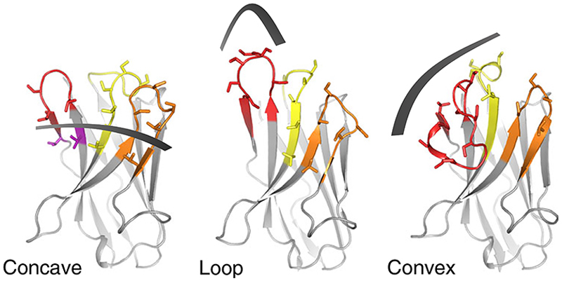
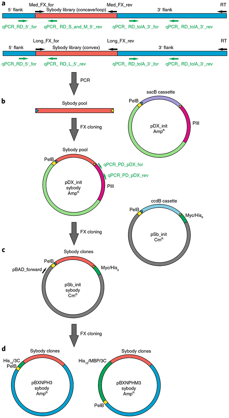
Here, we provide a protocol to generate synthetic nanobodies, known as sybodies, against any purified protein or protein complex within a 3-week period. Unlike methods that require animals for antibody generation, sybody selections are carried out entirely in vitro under controlled experimental conditions. This is particularly relevant for the generation of conformation-specific binders against labile membrane proteins or protein complexes and allows selections in the presence of non-covalent ligands. Sybodies are especially suited for cases where binder generation via immune libraries fails due to high sequence conservation, toxicity or insufficient stability of the target protein. The procedure entails a single round of ribosome display using the sybody libraries encoded by mRNA, followed by two rounds of phage display and binder identification by ELISA. The protocol is optimized to avoid undesired reduction in binder diversity and enrichment of non-specific binders to ensure the best possible selection outcome. Using the efficient fragment exchange (FX) cloning method, the sybody sequences are transferred from the phagemid to different expression vectors without the need to amplify them by PCR, which avoids unintentional shuffling of complementary determining regions. Using quantitative PCR (qPCR), the efficiency of each selection round is monitored to provide immediate feedback and guide troubleshooting. Our protocol can be carried out by any trained biochemist or molecular biologist using commercially available reagents and typically gives rise to 10-30 unique sybodies exhibiting binding affinities in the range of 500 pM-500 nM.


Conflict of interest statement

Competing interests

The authors declare competing financial interests. I.Z., P.E., R.J.P.D. and M.A.S. are co-founders and shareholders of Linkster Therapeutics AG.

Figures

Fig. 1
Fig. 1. Sybody libraries.
Shape and randomization scheme of the three sybody libraries: concave, loop and convex. CDR1, CDR2 and CDR3 are colored in yellow, orange and red, respectively. Randomized residues are depicted as sticks. A detailed description of the library design can be found in Zimmermann et al..
Fig. 2
Fig. 2. Sybody selection flowchart.
Sybody selections against target proteins start with one round of ribosome display, followed by two rounds of phage display. Binder hits are identified by ELISA and finally purified. The entire procedure is completed within a period of 3 weeks. PIII, filamentous phage protein III; α-Myc, antibody recognizing Myc-tag; Strep-HRP, streptavidin-horseradish peroxidase conjugate.
Fig. 3
Fig. 3. Overview of genetic constructs and primers.
a, The sybody libraries can be obtained from the authors in the form of mRNA ready for ribosome display. The concave and loop sybodies share the same framework and thus can be amplified with the same set of primers. The convex sybodies have a different framework. Primers used to quantify cDNA after ribosome display by qPCR (green) and to amplify the sybody pools by PCR (black) are indicated. b, Sybody pools amplified in a are cloned into the phagemid vector pDX_init using FX cloning. Note that the BspQI restriction sites (blue and yellow arrowheads) are encoded on the pDX_init backbone, allowing excision of the sybody open reading frames again. Primers used to quantify phages via pDX_init by qPCR are indicated in green. c, For single-clone expression, the output of the second phage display round is sub-cloned into pSb_init using FX cloning. Thereby, a Myc-tag and a His6-tag are attached at the C terminus of the sybodies. Note that the BspQI restriction sites are encoded on the pSb_init backbone. The sequencing primer pBAD_forward is indicated. d, For the production of tag-free proteins, sybodies are sub-cloned into pBXNPH3 or pBXNPHM3. In this cloning step, the BspQI restriction sites are lost.
Fig. 4
Fig. 4. Exemplary DNA gel of sybody pools.
The entire PCR reaction (100 µl) obtained from amplifying cDNA encoding for enriched pools of the concave, loop and convex sybodies was loaded on a preparative 1.5% (wt/vol) agarose gel in 1× TEA buffer and stained with ethidium bromide (Step 22). The length differences of the three sybody libraries can be distinguished (concave: 342 bp; loop: 360 bp; convex: 372 bp).
Fig. 5
Fig. 5. SEC analysis of sybodies.
Three exemplary sybodies were purified from medium-scale cultures and analyzed on a Sepax SRT-10C SEC100 column. The sybody giving rise to the blue chromatogram elutes at a retention volume of ~11 ml and is thus a monomer. The red chromatogram with its peak <10 ml represents an oligomerizing sybody. The orange chromatogram with its main peak at ~16 ml represents a sybody exhibiting strong column interaction. AU, arbitrary units.
Fig. 6
Fig. 6. A conformation-specific sybody against the ABC transporter TM287/288.
a, The convex sybody Sb_TM#35 (gray with CDR3 in red) served as a crystallization chaperone to solve the structures of outward-facing ABC exporter TM287/288 (TM287 chain in aquamarine, TM288 chain in light pink, bound ATP as spheres) (PDB: 6QUZ). b, SPR analysis showing that the sybody Sb_TM#35 binds only to the outward-facing ABC transporter in the presence of ATP and Mg2+. KD, dissociation constant; RU, response units. The data shown in b were taken from ref. .
Fig. 7
Fig. 7. Structure of the KDEL receptor in complex with a sybody.
a, The loop sybody Syb37 occupies the KDEL peptide-binding pocket of the KDEL receptor via its CDR3 sequence (red) (PDB: 6I6J). b, Peptide-binding assays using tritiated TAEKDEL peptide showing that sybody Syb37 competes with peptide binding. The data shown in panel b were taken from ref. 7. Column heights indicate mean values and the error bars are standard deviations of the individual measurement points shown as circles.

References

    1. Ries J, Kaplan C, Platonova E, Eghlidi H, Ewers H. A simple, versatile method for GFP-based super-resolution microscopy via nanobodies. Nat Methods. 2012;9:582–584. - PubMed
    1. Rothbauer U, et al. Targeting and tracing antigens in live cells with fluorescent nanobodies. Nat Methods. 2006;3:887–889. - PubMed
    1. Manglik A, Kobilka BK, Steyaert J. Nanobodies to study G protein-coupled receptor structure and function. Annu Rev Pharmacol Toxicol. 2017;57:19–37. doi: 10.1146/annurev-pharmtox-010716-104710. - DOI - PMC - PubMed
    1. Harmansa S, Alborelli I, Bieli D, Caussinus E, Affolter M. A nanobody-based toolset to investigate the role of protein localization and dispersal in Drosophila. Elife. 2017;6:e22549. doi: 10.7554/eLife.22549. - DOI - PMC - PubMed
    1. Bukowska MA, Grutter MG. New concepts and aids to facilitate crystallization. Curr Opin Struct Biol. 2013;23:409–416. - PubMed

Publication types

MeSH terms

Substances

LinkOut - more resources