. 2020 Mar 15;12(3):813-824.
eCollection 2020.

Identification of differentially expressed microRNAs and their target genes in the hippocampal tissues of Fmr1 knockout mice

Affiliations

Identification of differentially expressed microRNAs and their target genes in the hippocampal tissues of Fmr1 knockout mice

Malan Zhang et al. Am J Transl Res. .

Abstract

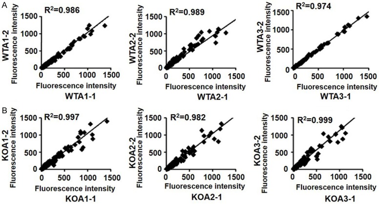
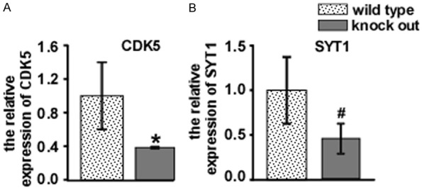
Fragile X syndrome (FXS) is one of the most common forms of inherited mental retardation; it is usually associated with the transcriptional silencing of the Fmr1 gene and loss of its encoded protein, the fragile X mental retardation protein (FMRP). FMRP is an RNA-binding protein and participates in regulating the development of dendritic spines and synaptic plasticity. To uncover the possible role of microRNAs (miRNAs) in FXS and their relationship with FMRP, we used microarray analysis to investigate the miRNA expression profiles in the hippocampal tissues of Fmr1 knockout (Fmr1-KO) mice and wild type (WT) mice. A total of 75 differentially expressed miRNAs were identified, of which 58 were significantly upregulated and no miRNAs were significantly downregulated in Fmr1-KO mice. Quantitative real-time PCR (qRT-PCR) analysis was applied to validate the expression of 7 upregulated miRNAs; results indicated that the levels of only miR-449a and miR-720 were significantly upregulated. We further used bioinformatics software and databases to predict the target genes of these two miRNAs. The genes were related to dendritic spine development and synaptic plasticity; the qRT-PCR and western blotting results showed that cyclin-dependent kinase 5 (CDK5) and synaptotagmin 1 (SYT1) were differentially expressed in the Fmr1-KO mice and WT mice. In conclusion, this study evidenced diverse changes in the expression of miRNAs, and validated the miRNAs and their targeted genes in Fmr1-KO mice. Although further studies are required to better understand the function of miRNAs in FXS, the present research highlights a potential role of miRNAs in the pathogenesis of FXS.

Keywords: Fragile X syndrome; fragile X mental retardation protein; microRNAs; microarray analysis; synaptic plasticity.


Conflict of interest statement

None.

Figures

Figure 1
Figure 1
The FMRP protein, which is the expression product of the Fmr1 gene, was absent in the Fmr1 knockout mice but present in the wild type mice. Western blotting was performed to validate the FMRP protein levels in the Fmr1 knockout mice and wild type mice. WT1 and WT2 indicate the wild type animal 1 and wild type animal 2, respectively. KO1 and KO2 indicate the knockout animal 1 and knockout animal 2, respectively.
Figure 2
Figure 2
The experimental repeatability of the microarray analysis was between 0.974 and 0.999. WTA1, WTA2, and WTA3 indicate the wild type animal 1, wild type animal 2, and wild type animal 3, respectively. KOA1, KOA2, and KOA3 indicate the knockout animal 1, knockout animal 2, and knockout animal 3, respectively.
Figure 3
Figure 3
Changes of the miRNA expression in the hippocampal tissues of the Fmr1 knockout mice and wild-type mice. The heatmap shows the hierarchical clustering of miRNAs in each animal from the Fmr1 knockout and wild type groups. The yellow color indicates that the miRNA expression levels in the samples were upregulated, and the blue color indicates that the miRNA expression levels in the samples were downregulated. All differentially expressed miRNAs are listed in the Supplementary Tables 3 and 4.
Figure 4
Figure 4
The relative expression of miR-720 and miR-449a in the hippocampal tissues of the Fmr1 knockout mice was higher than that in the hippocampal tissues of the wild type mice. QRT-PCR was performed to validate the miR-720 and miR-449a expression levels in the Fmr1 knockout mice and wild type mice. The miRNA expression levels in the mice from the Fmr1 knockout group were normalized to those in the mice from the wild-type group. *P < 0.01, #P < 0.05.
Figure 5
Figure 5
The CDK5 and SYT1 mRNA levels in the hippocampal tissues of the Fmr1 knockout mice were lower than those in the hippocampal tissues of the wild type mice. qRT-PCR was performed to validate the CDK5 and SYT1 mRNA expression levels in the Fmr1 knockout mice and wild type mice. The mRNA expression levels in the mice from the Fmr1 knockout group were normalized to those in the mice from the wild-type group. *P < 0.01, #P < 0.05.
Figure 6
Figure 6
The CDK5 and SYT1 protein levels in the hippocampal tissues of the Fmr1 knockout mice were lower than those in the hippocampal tissues of the wild type mice. Western blotting was performing to validate the CDK5 and SYT1 protein levels in the Fmr1 knockout mice and wild type mice. The protein levels in the mice from the Fmr1 knockout group were normalized to those in the mice from the wild-type group. *P < 0.01, #P < 0.05.

References

    1. Pfeiffer BE, Huber KM. The state of synapses in fragile X syndrome. Neuroscientist. 2009;15:549–567. - PMC - PubMed
    1. Bassell GJ, Warren ST. Fragile X syndrome: loss of local mRNA regulation alters synaptic development and function. Neuron. 2008;60:201–214. - PMC - PubMed
    1. Basehore MJ, Friez MJ. Molecular analysis of fragile X syndrome. Curr Protoc Hum Genet. 2014;80 Unit 9.5. - PubMed
    1. Sethna F, Moon C, Wang H. From FMRP function to potential therapies for fragile X syndrome. Neurochem Res. 2014;39:1016–1031. - PMC - PubMed
    1. Najib A, Kim MS, Choi SH, Kang YJ, Kim KH. Changes in microRNAs expression profile of olive flounder (paralichthys olivaceus) in response to viral hemorrhagic septicemia virus (VHSV) infection. Fish Shellfish Immunol. 2016;51:384–391. - PubMed

LinkOut - more resources