Review
. 2020 Mar 31:9:F1000 Faculty Rev-222.
doi: 10.12688/f1000research.20576.1. eCollection 2020.

Emerging concepts in ventilation-induced lung injury

Affiliations
Review

Emerging concepts in ventilation-induced lung injury

Purnema Madahar et al. F1000Res. .

Abstract

Ventilation-induced lung injury results from mechanical stress and strain that occur during tidal ventilation in the susceptible lung. Classical descriptions of ventilation-induced lung injury have focused on harm from positive pressure ventilation. However, injurious forces also can be generated by patient effort and patient-ventilator interactions. While the role of global mechanics has long been recognized, regional mechanical heterogeneity within the lungs also appears to be an important factor propagating clinically significant lung injury. The resulting clinical phenotype includes worsening lung injury and a systemic inflammatory response that drives extrapulmonary organ failures. Bedside recognition of ventilation-induced lung injury requires a high degree of clinical acuity given its indistinct presentation and lack of definitive diagnostics. Yet the clinical importance of ventilation-induced lung injury is clear. Preventing such biophysical injury remains the most effective management strategy to decrease morbidity and mortality in patients with acute respiratory distress syndrome and likely benefits others at risk.

Keywords: acute lung injury; acute respiratory distress syndrome; mechanical ventilation; respiratory mechanics; ventilator-induced lung injury.


Conflict of interest statement

No competing interests were disclosed.No competing interests were disclosed.No competing interests were disclosed.No competing interests were disclosed.No competing interests were disclosed.

Figures

Figure 1.
Figure 1.. Local stress and strain of epithelial cells generated during recruitment of small airways.
( A) Air bubble propagation down the atelectatic airway generates a dynamic wave of stress and strain at the interface of the air bubble and collapsed airway. As the air bubble approaches, the epithelial cell is pulled inward toward the bubble. As the air bubble passes, the cell is pushed outward. ( B) The air bubble similarly generates stress and strain of epithelial cells during propagation along flooded airway. This figure was re-used from Ghadiali SN and Gaver DP, Biomechanics of liquid–epithelium interactions in pulmonary airways. Respir Physiol Neurobiol doi:10.1016/j.resp.2008.04.008 with permission .
Figure 2.
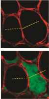
Figure 2.. Spatial heterogeneity increases stress and strain due to alveolar interdependence.
In the isolated perfused rat lung, confocal microscopy with optical sections 2 µm thick permits direct visualization of alveoli. Top: adjacent alveoli share a common septum and are mechanically interdependent. In normal conformation, strain is minimized across neighboring air-filled alveoli. Bottom: in a single-alveolus model of pulmonary edema, the effects on local strain distribution of heterogeneous parenchymal consolidation and flooding can be appreciated. The liquid-filled alveolus shrinks owing to micromechanical effects of meniscus formation. As a result, the adjacent air-filled alveolus bulges and overdistends. This figure was reprinted with permission of the American Thoracic Society. Copyright © 2020 American Thoracic Society. Cite: Perlman CE, Lederer DJ, Bhattacharya J. 2011. Micromechanics of alveolar edema. Am J Respir Cell Mol Biol, 44(1), 34–9. The American Journal of Respiratory Cell and Molecular Biology is an official journal of the American Thoracic Society .
Figure 3.
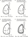
Figure 3.. Transpulmonary pressure (Pairway – Ppleural) is the pertinent distending pressure of the lung.
Measurements to ascertain lung distension at end-inspiration and end-expiration are taken during breath holds, at which time air flow is zero and airway pressure equilibrates throughout communicating airways. ( A) Lean, non-intubated patient with normal spontaneous tidal breathing at end inspiration. Transpulmonary pressure of 5–10 cmH 2O is typical at end inspiration in lean healthy individuals. ( B) Lean, intubated patient during positive pressure ventilation at end inspiration while passive. ( C) Lean, intubated patient with forceful inspiratory effort has produced very high transpulmonary pressure that would be unsafe in patients at risk of ventilation-induced lung injury (VILI) despite the relatively low airway pressure. The observed transpulmonary pressure of 25 cmH 2O is typical at total lung capacity in lean healthy individuals. ( D) Obese, intubated patient, with chest wall contributing high pleural pressure that results in lower transpulmonary pressure and lower lung volume at end inspiration despite higher airway pressure. Even though plateau airway pressure is relatively high, the risk of VILI is lower because lung volume and transpulmonary pressure are lower. Paw, airway pressure; Ppl, pleural pressure; Ptp, transpulmonary pressure. This figure was adapted from Beitler J, Malhotra A, and Thompson B, Ventilator-induced lung injury. Clin Chest Med doi: 10.1016/j.ccm.2016.07.004 with permission .

Similar articles

Cited by

References

    1. Amato MB, Barbas CS, Medeiros DM, et al. : Effect of a protective-ventilation strategy on mortality in the acute respiratory distress syndrome. N Engl J Med. 1998;338(6):347–54. 10.1056/NEJM199802053380602 - DOI - PubMed
    1. Acute Respiratory Distress Syndrome Network, Brower RG, Matthay MA, et al. : Ventilation with lower tidal volumes as compared with traditional tidal volumes for acute lung injury and the acute respiratory distress syndrome. N Engl J Med. 2000;342(18):1301–8. 10.1056/NEJM200005043421801 - DOI - PubMed
    2. F1000 Recommendation

    1. Slutsky AS, Ranieri VM: Ventilator-induced lung injury. N Engl J Med. 2013;369(22):2126–36. 10.1056/NEJMra1208707 - DOI - PubMed
    1. Matthay MA, Zemans RL, Zimmerman GA, et al. : Acute respiratory distress syndrome. Nat Rev Dis Primers. 2019;5(1):18. 10.1038/s41572-019-0069-0 - DOI - PMC - PubMed
    1. Lanspa MJ, Gong MN, Schoenfeld DA, et al. : Prospective Assessment of the Feasibility of a Trial of Low Tidal Volume Ventilation for Patients with Acute Respiratory Failure. Ann Am Thorac Soc. 2019;16(3):356–362. 10.1513/AnnalsATS.201807-459OC - DOI - PMC - PubMed
    2. F1000 Recommendation

Publication types

LinkOut - more resources