

Review
doi: 10.12688/f1000research.22405.1.
eCollection 2020.
Astrocytogenesis: where, when, and how
Affiliations
- PMID: 32269761
- PMCID: PMC7122459
- DOI: 10.12688/f1000research.22405.1
Item in Clipboard
Review
Astrocytogenesis: where, when, and how
F1000Res.
.
Abstract
Astrocytes are the most abundant cell type in the central nervous system and have diverse functions in blood-brain barrier maintenance, neural circuitry formation and function, and metabolic regulation. To better understand the diverse roles of astrocytes, we will summarize what is known about astrocyte development and the challenges limiting our understanding of this process. We will also discuss new approaches and technologies advancing the field.
Keywords: Astrocyte; Glia; Gliogenesis; Neurodevelopment.
Copyright: © 2020 Akdemir ES et al.
Conflict of interest statement
No competing interests were disclosed.No competing interests were disclosed.No competing interests were disclosed.
Figures
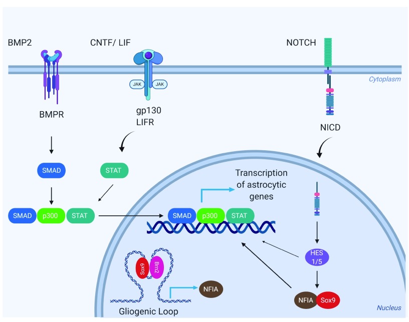
Neural stem cells (NSCs) express astrocytic genes in response to several signaling molecules, including bone morphogenic protein (BMP) families, the leukemia inhibitory factor/ciliary neurotrophic factor (LIF/CNTF), and Notch pathway. BMPs signal primarily through SMAD, whereas LIF/CNTF activates the JAK/STAT pathways. The active SMAD–STAT complex bridged by p300 goes directly into the nucleus, binds to the promoter, and activates astrocytic genes such as
GFAP and
S100. Another important pathway that regulates astrocytogenesis is the Notch pathway. Notch ligands will activate Notch receptors and activate the expression of Hes genes,
Hes1/5. Hes1, Hes5, and activated forms of Notch receptors induce the expression of astrocytic genes and glial promoting transcription factor nuclear factor-I A (NFIA). In early development, a pre-formed gliogenic loop serves to facilitate the association between sex-determining region Y-box 9 (Sox9) and brain-specific homeobox/POU domain protein 2 (Brn2), which also drives expression of NFIA. NFIA is both necessary and sufficient for the induction of astrocytic genes.
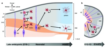
(
a) In the developing cortex, radial glia (RG) first give rise to glioblasts (GBs) during the late embryonic to perinatal period. Glioblasts undergo several rounds of division while migrating out along radial glia, resulting in clusters of astrocytes in the developing cortex. At the terminal stage of radial glial differentiation, radial glia detach from the ventricular zone and form unipolar transitional radial glia (tRG), which give rise to protoplasmic and fibrous astrocytes in the gray matter and white matter of the cortex, respectively. During the early postnatal period, differentiated astrocytes in the outer cortical layer undergo symmetric division and generate daughter astrocytes that exhibit astrocytic morphology and functions. (
b) In the developing spinal cord, radial glia first proliferate during embryonic day 12 (E12) to 13, giving rise to radial glial pool which differentiates into astrocytes between E14 and postnatal day 3 (P3). Alternatively, radial glial cells differentiate into intermediate astrocyte precursors (IAPs), which proliferate during E14 to P3 and undergo terminal differentiation, ultimately giving rise to astrocytes. The progression from embryonic stage to adult is shown from left to right below each panel. Straight arrows indicate differentiation or maturation from one cell type to another. Circular arrows indicate proliferation. Dashed arrows indicate migration. CP, cortical plate; fAstro, fibrous astrocyte; GM, gray matter; iAstro, immature astrocyte; P, pia mater; pAstro, protoplasmic astrocyte; SVZ, subventricular zone; VZ, ventricular zone; WM, white matter.
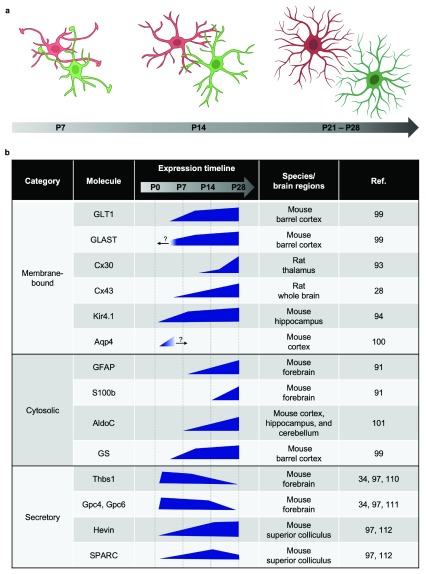
(
a) Morphology of astrocytes matures in a stepwise manner. At postnatal day 7 (P7), astrocytes exhibit few branches and the overall morphology is simple and territories are small. Long protrusions are often seen extending into the territories of the neighboring astrocytes. The boundaries of territories are not well-defined yet. At postnatal day 14 (P14), the number of branches become greater and astrocyte territories become bigger. More branch points grow out of existing branches, resulting in more complex morphology. Long protrusions are seen less, and the boundaries of territories start to emerge. At postnatal day 21 to day 28 (P21–P28), astrocytes develop their characteristic “spongiform” complex morphology, and there is minimal overlap between neighboring astrocytes and clear boundary of each territory. (
b) Genes that are induced during astrocyte maturation. Blue polygons indicate expression levels of the genes during development. Question marks indicate that the expression level beyond the developmental stage is unknown.
References
Publication types
MeSH terms
LinkOut - more resources
Full Text Sources