. 2020 Mar 18:8:157.
doi: 10.3389/fcell.2020.00157. eCollection 2020.

Human Derived Immortalized Dermal Papilla Cells With a Constant Expression of Testosterone Receptor

Affiliations

Human Derived Immortalized Dermal Papilla Cells With a Constant Expression of Testosterone Receptor

Tomokazu Fukuda et al. Front Cell Dev Biol. .

Abstract

Androgenetic alopecia (AGA) is the most common type of hair loss, and is mainly caused by the biological effects of testosterone on dermal papilla cells (DPCs). In vitro culturing of DPCs might be a useful tool for the screening of target molecule of AGA. However, primary DPCs cannot continuously proliferate owing to cellular senescence and cell culture stress. In this study, we introduced mutant cyclin-dependent kinase 4 (CDK4), Cyclin D1, and telomerase reverse transcriptase (TERT) into DPCs. We confirmed protein expression of CDK4 and Cyclin D1, and enzymatic activity of TERT. Furthermore, we found the established cell line was free from cellular senescence. We also introduced the androgen receptor gene using a recombinant retrovirus, to compensate the transcriptional suppressed endogenous androgen receptor in the process of cell proliferation. Furthermore, we detected the efficient nuclear translocation of androgen receptor into the nucleus after the treatment of dihydrotestosterone, indicating the functionality of our introduced receptor. Our established cell line is a useful tool to identify the downstream signaling pathway, which activated by the testosterone.

Keywords: androgen receptor; dermal papilla cells; dihydrotestosterone; immortalization; nuclear localization.


Figures

FIGURE 1
FIGURE 1
The morphologies of wild type, EGFP-expressing, mutant CDK4 and Cyclin D (K4D cell)-expressing, and mutant CDK4, Cyclin D, and telomere reverse transcriptase (TERT)-expressing cells (K4DT cells). (A) Cell morphology of wild type (upper left), EGFP (upper right), CDK4 and Cyclin D (lower left), and mutant CDK4, Cyclin D, and TERT-expressing cells. (B) Detection of EGFP fluorescence after infection of DPCs with EGFP-expressing lentiviruses. Fluorescent, difference in contrast (DIC), and merge images are shown.
FIGURE 2
FIGURE 2
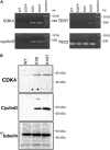
Detection of integration of the expression cassette into DPC genomes with PCR, and detection of protein expression of the introduced genes. (A) PCR detection of the expression cassettes of CDK4, Cyclin D, and TERT in the genomic DNA of DPCs. PCR products from Tuberous sclerosis type 2 (TSC2) were used as a control. (B) Western blot analysis of wild type, K4D, and K4DT cells. The results obtained from CDK4, Cyclin D, and α-tubulin antibodies are shown.
FIGURE 3
FIGURE 3
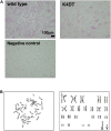
Detection of the alkaline phosphatase activity and the chromosome conditions of established DPCs. (A) Detection of alkaline phosphatase activity using Fast Red in wild type (upper left), K4DT (upper right), and rat-derived fibroblasts (lower right, negative control). (B) Chromosome analysis of K4DT cells. Left panel, a representative mitotic chromosome pattern from a K4DT cell. Right panel, an aligned G-banding chromosome pattern obtained from a K4DT cell. Sexual chromosomes are labeled as X and Y.
FIGURE 4
FIGURE 4
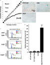
Results of sequential passaging, detection of cellular senescence, and expression levels of the androgen receptor. (A) Sequential passaging and cumulative population doubling of wild type, K4D, and K4DT cells. Averages and standard deviations were obtained from triplicate samples. (B) Detection of cellular senescence using SA-β-gal staining in wild type, K4D, and K4DT cells. The bar indicates 100 μm. (C) Cell cycle histogram of representative results obtained from wild type, K4D, and K4DT cells. (D) Detection of the androgen receptor gene in wild type DPCs at passage 8, immortalized K4DT DPCs at passage 12, HE12 human fibroblasts, and adult human prostate-derived RNA. Wild type DPC expression levels were set to 1.0, and the relative amount of AR was detected using real-time PCR. Six samples were analyzed for each group. The averages and standard deviations are shown.
FIGURE 5
FIGURE 5
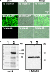
Retroviral introduction of the androgen receptor into immortalized DPCs with K4DT. (A) G418 selection of wild type DPCs (no infection), QCXIN-EGFP-infected DPCs, and QCXIN-AR-infected DPCs. Note that wild type (no infection) cells exhibited complete cell death upon addition of 700 μg/mL G418. (B) Western blot analysis of QCXIN-EGFP (lane 1) and QCXIN-AR (lane 2) probed with HA protein tag antibody (Left panel), and α-tubulin antibody (Right panel). Note that the HA positive band was observed at around 100 kDa in the left panel.
FIGURE 6
FIGURE 6
Immunohistochemical detection of the androgen receptor using an HA antibody. (A) Immunohistochemical detection of androgen receptor-expressing DPCs immortalized with expression of mutant CDK4, Cyclin D, and TERT. EGFP (left panels), androgen receptor detected using an HA antibody (middle panels), and a merged image with EGFP, androgen receptor staining, and nuclear counterstaining with DAPI (Right panels). Upper panels are low magnification. Lower panels are high magnification. Note that nuclear and cytoplasmic localization of androgen receptor detected with HA antibody. (B) Detection of fluorescence in immortalized DPCs expressing QCXIN-EGFP. EGFP fluorescence (Right panels), HA antibody staining (middle panels), and a merged image with EGFP, HA antibody staining, and nuclear counterstaining with DAPI (Right panels). Lower panels are high magnification.
FIGURE 7
FIGURE 7
Detection of cytoskeletal F-actin in wild type, K4DT, AR expressing K4DT DPCs. (A) Staining feature of wild type, K4DT, and AR expressing K4DT DPCs. Staining with phalloidin, DAPI, and merged pictures were presented. (B) The quantitation of fluorescence intensity from the cytoplasm of 15 randomly selected cells. Average and standard error were shown in the graph. No significant difference.
FIGURE 8
FIGURE 8
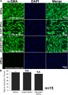
Detection of α-smooth muscle actin (SMA) in wild type, K4DT, AR expressing K4DT DPCs. (A) Staining feature of wild type, K4DT, and AR expressing K4DT DPCs. Note that there is no staining signal of SMA in the absence of primary antibody. Staining signals of SMA, DAPI, and Merged pictures were shown. (B) The quantitation of fluorescence intensity from the cytoplasm of 15 randomly selected cells. Average and standard error were shown in the graph. No significant difference.
FIGURE 9
FIGURE 9
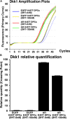
Detection of Dkk1 expression before and after the exogeneous introduction of androgen receptor (AR), and absence and presence of dihydrotestosterone (DHT). (A) Amplification plots of endogenous Dkk1 in EGFP expressing K4DT DPCs and AR expressing K4DT DPCs. (B) The quantitation of Dkk1 expression with ΔΔ Ct method. The relative quantitation value of samples were shown in the graph. When one of the sample of EGFP expressing K4DT DPCs was set as 1.0, the expression level of samples were calculated after the adjustment with glyceraldehyde-3-phosphate dehydrogenase (GAPDH). Number of samples is 6 for each group.
FIGURE 10
FIGURE 10
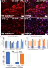
Detection of nuclear translocation of androgen receptor (AR) under absence and presence of dihydrotestosterone (DHT). (A) Representative staining feature of AR K4DT DPCs with and without DHT. Note that under the presence of DHT, the localization of AR in the cytoplasm is almost negative, and AR localize within the nuclear. (B) Count of cells, which positive nuclear and cytoplasm (blue bar) and cells, which positive only within the nuclear (orange bar). Left side, results from 16 images before DHT treatment. Right side, results from 16 images after DHT treatment. Bottom panel, percentage of cells, which positive both nuclear and cytoplasm (blue bar) and percentage of cells, which positive only within the nuclear (orange bar). The statistical significance more than 0.1% were shown with three stars. N = 16 for each group.
FIGURE 11
FIGURE 11
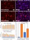
Nuclear translocation of AR by DHT is partially inhibited by MVD3100 in AR expressing K4DT DPCs. (A) Representative staining feature of AR with HA tag antibody after the DHT treatment, under the absence and presence of MDV3100. (B) Upper panel, Count of cells, which positive nuclear and cytoplasm (blue bar) and cells, which positive only within the nuclear (orange bar) under the absence of MDV3100. Lower panel, Count of cells, which positive nuclear and cytoplasm (blue bar) and cells, which positive only within the nuclear (orange bar) under the existence of MDV3100. (C) Percentage of cells, which positive both nuclear and cytoplasm (blue bar) and percentage of cells, which positive only within the nuclear (orange bar). Comparison between absence of presence of MDV3100. The statistical significance more than 0.1% were shown with three stars. N = 16 for each group.

References

    1. Coskuner E. R., Ozkan B., Culha M. G. (2019). Sexual problems of men with androgenic alopecia treated with 5-Alpha reductase inhibitors. Sex. Med. Rev. 7 277–282. 10.1016/j.sxmr.2018.07.003 - DOI - PubMed
    1. Danilenko D. M., Ring B. D., Pierce G. F. (1996). Growth factors and cytokines in hair follicle development and cycling: recent insights from animal models and the potentials for clinical therapy. Mol. Med. Today 2 460–467. 10.1016/1357-4310(96)10045-9 - DOI - PubMed
    1. Davis B. K. (1962). Phases of the hair-growth cycle. Nature 194 694–694. 10.1038/194694a0 - DOI - PubMed
    1. Debacq-Chainiaux F., Erusalimsky J. D., Campisi J., Toussaint O. (2009). Protocols to detect senescence-associated beta-galactosidase (SA-βgal) activity, a biomarker of senescent cells in culture and in vivo. Nat. Protoc. 4 1798–1806. 10.1038/nprot.2009.191 - DOI - PubMed
    1. Detmar M., Schaart F. M., Blume U., Orfanos C. E. (1993). Culture of hair matrix and follicular keratinocytes. J. Invest. Dermatol. 101 130S–134S. 10.1016/0022-202X(93)90513-H - DOI - PubMed