Review
. 2020 Mar 25:8:190.
doi: 10.3389/fcell.2020.00190. eCollection 2020.

The Influence of Neuron-Extrinsic Factors and Aging on Injury Progression and Axonal Repair in the Central Nervous System

Affiliations
Review

The Influence of Neuron-Extrinsic Factors and Aging on Injury Progression and Axonal Repair in the Central Nervous System

Theresa C Sutherland et al. Front Cell Dev Biol. .

Abstract

In the aging western population, the average age of incidence for spinal cord injury (SCI) has increased, as has the length of survival of SCI patients. This places great importance on understanding SCI in middle-aged and aging patients. Axon regeneration after injury is an area of study that has received substantial attention and made important experimental progress, however, our understanding of how aging affects this process, and any therapeutic effort to modulate repair, is incomplete. The growth and regeneration of axons is mediated by both neuron intrinsic and extrinsic factors. In this review we explore some of the key extrinsic influences on axon regeneration in the literature, focusing on inflammation and astrogliosis, other cellular responses, components of the extracellular matrix, and myelin proteins. We will describe how each element supports the contention that axonal growth after injury in the central nervous system shows an age-dependent decline, and how this may affect outcomes after a SCI.

Keywords: aging; astrocytes; extra-cellular matrix; inflammation; microglia; neuron-extrinsic factors; signaling; spinal cord injury.


Figures

FIGURE 1
FIGURE 1
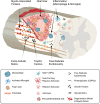
Summary schematic of the cellular and extracellular cellular factors that can influence axon growth in the chronic spinal cord injury scar and that may be altered with age. In response to traumatic spinal injury, many cellular and molecular mechanisms are activated, including an inflammatory response (neutrophils, monocytes, microglia, macrophages), astrocytic response (forming the astroglial scar), and fibroblast response (from different origins, forming the fibrotic scar). With age, the initial injury response is altered and a reduction of locomotor functional recovery is observed. There is more BSCB permeability (resulting in more blood cells and molecules entering the injury), more pro-inflammatory response (more “M1-phenotype”), reduction in wound healing (linked to the alteration in the astrocytic response), induction of cell death (including neurons and oligodendrocytes). In the more chronic phase of the injury, age keeps changing the cellular response compared to young animals (inflammatory cells, astrocytes, fibroblasts, OPCs, endothelial, pericytes) which alters the environment: there is more debris present (including axon and myelin debris), alteration in the growth factors produced, changes in ECM composition (changes in glycoproteins and proteoglycans production), and less revascularization. All of these can participate in the age-dependent decline in axon regeneration. BSCB, blood spinal cord barrier; ECM, extracellular matrix; OPCs, oligodendrocytes precursor cells.
FIGURE 2
FIGURE 2
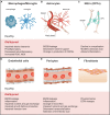
The cellular contributors to spinal cord injury altered in aging further reduce injury repair and axon growth. (A) Inflammatory response by microphages and microglia results in more a pro-inflammatory and less anti-inflammatory profile, associated with a reduction in debris clearance efficiency, and increase in ROS and NOX. (B) Astrocytes form the astroglial scar and their alteration with age and injury may include changes in ECM molecule expression profiles (growth factors, glycoproteins, proteoglycan production - CSPGs). Astrocytes become pro-inflammatory, resulting in increases in ROS and NOX. Changes in astrocyte functions also include increases in BSBC leakiness and reduction in nutrient availability. (C) Fibroblasts, from different origins, form the fibrotic scar; with age and injury, the profile of the ECM molecules they produce changes, making the injury more anti-regenerative, altering the injury stiffness, and making a physical barrier to growth. (D) Changes in the state of the endothelial cells with age can lead to increases in BSBC leakiness, infiltration of inflammatory molecules, reduction in growth factor production, and reduction in control of the vascular tone, leading to local hypoxia and reduction in nutrients entry. (E) Similar to endothelial cells, alteration in the functions of pericytes with age and injury may lead to vascular dysfunction, reduction in trophic support, increase in local hypoxia and decrease in angiogenesis. The roles of pericytes in the fibrotic scar and production of ECM molecules may also be altered. (F) With age, the proliferation and differentiation potential of OPCs is reduced, which can diminish the potential for remyelination, alter neuroinflammation and reduce neuron survival after SCI. These changes can also alter the astrocytic scar, making it more inhibitory. BSCB, blood spinal cord barrier; CSPGs, chondroitin sulfate proteoglycans; ECM, extracellular matrix; OPCs, oligodendrocytes precursor cells; NOX, NADPH-oxidase; ROS, reactive oxygen species.
FIGURE 3
FIGURE 3
Alterations in microglia in aging, and their responses to spinal cord injury, exacerbate the injury and reduce axon growth. (A) Young Naïve Microglia. In the young naïve CNS, resting microglia have a ramified appearance with branched processes, and have an organized distribution. These cells are involved in extracellular surveillance, debris clearance, environmental maintenance, and antigen presentation. (B) Young Activated Microglia. In the young CNS, microglia are activated after a SCI, becoming more ameboid and phagocytic. This is characterized by increased debris clearance, upregulation of inflammatory mediators (mostly pro- and some anti-inflammatory), activation of inflammatory signaling pathways, and the production of ROS and RNS. (C) Aged Naïve Microglia. In the aging CNS, microglia undergo physiological and functional changes linked with a systematic para-inflammatory state. Aged microglia have a de-ramified appearance and a more disorganized distribution. These cells are less plastic and become ‘primed,’ characterized by increased expression of pro-inflammatory mediators, increased NOX, ROS and RNS, impaired debris clearance, and liposome accumulation. (D) Aged Activated Microglia. After a SCI in the aged CNS, aged microglia respond similarly to the younger microglia. However, these aged cells are already ‘primed,’ pro-inflammatory, and have impaired debris clearance. This results in exacerbation of the detrimental elements of microglial activation, such as excessive production of pro-inflammatory mediators into the lesion environment, excessive NOS, ROS and RNS leading to increased oxidative stress, and inefficient debris clearance. CNS, Central Nervous System; NOX:, NADPH-oxidase; ROS, Reactive Oxygen Species; RNS, Reactive Nitrogen Species; SCI, Spinal Cord Injury.
FIGURE 4
FIGURE 4
Differences in astrocyte phenotypes, functions and responses with aging play a significant role in central nervous system health as well as spinal cord injury response. (A) Regular Function of Young Astrocytes. Astrocytes play a vital role in cellular, molecular, and metabolic homeostasis in the naïve CNS. These cells are involved in synaptogenesis and support mature synapses. They also support neurons and neuronal function through the uptake of glutamate, K+, and GABA, the production and release of transmitters, cytokines and ATP, and by stocking glycogen. Astrocyte processes interact with vascular endothelial cells and are an important element of the BBB. (B) Young Reactive Astrocytes. After SCI, astrocytes become reactive, increasing in size and branching. These reactive astrocytes are characterized by a suite of molecular changes including increased GFAP expression, increased proteoglycan (CSPGs) production, increased NOS and ROS, and production of inflammatory cytokines. Reactive astrocytes are involved in BBB repair, and sequestering the inflammatory lesion and fibrotic scar. (C) Normal Aging Astrocytes. In the aging CNS, astrocytes become larger and more branched than their younger un-injured counterparts. These cells exhibit an early stage of reactive astrogliosis characterized by increased GFAP, vimentin and S100β expression, increased expression of growth factors (such as CNTF and TGF-β) and increased oxidoreductive enzymes and oxidative stress. These cells show impaired ability to perform normal functions such as BBB/vascular maintenance and neuronal support. (D) Aged Reactive Astrocytes. After SCI, the alterations in astrocytes with aging are exacerbated. Aged astrocytes are already in an early reactive state, this is compounded by injury and activation cues in the lesion environment. The result is greater increase in GFAP, increase in oxidative stress and inflammation, increased excitotoxicity, impaired BBB repair, and, significantly, impaired sequestering of the inflammatory lesion. All of this will have profound effects on neuronal survival and axon re-growth. BBB, Blood-Brain Barrier; CNS, Central Nervous System; CNTF, Ciliary Neurotrophic Factor; CSPGs, Chondroitin Sulfate Proteoglycans; GFAP, Glial Fibrillary Acidic Protein; NOS, Nitric Oxide Synthase; ROS, Reactive Oxygen Species; SCI, Spinal Cord Injury.

References

    1. Abbott N. J. (2002). Astrocyte–endothelial interactions and blood–brain barrier permeability. J. Anat. 200 629–638. 10.1046/j.1469-7580.2002.00064.x - DOI - PMC - PubMed
    1. Adams R. H., Eichmann A. (2010). Axon guidance molecules in vascular patterning. Cold Spring Harb. Perspect. Biol. 2:a001875. 10.1101/cshperspect.a001875 - DOI - PMC - PubMed
    1. Al-Shawi R., Hafner A., Chun S., Raza S., Crutcher K., Thrasivoulou C., et al. (2007). ProNGF, sortilin, and age-related neurodegeneration. Ann. N. Y. Acad. Sci. 1119 208–215. 10.1196/annals.1404.024 - DOI - PubMed
    1. Al-Shawi R., Hafner A., Olson J., Chun S., Raza S., Thrasivoulou C., et al. (2008). Neurotoxic and neurotrophic roles of proNGF and the receptor sortilin in the adult and ageing nervous system. Eur. J. Neurosci. 27 2103–2114. 10.1111/j.1460-9568.2008.06152.x - DOI - PubMed
    1. Anderson M. A., Ao Y., Sofroniew M. V. (2014). Heterogeneity of reactive astrocytes. Neurosci. Lett. 565 23–29. 10.1016/j.neulet.2013.12.030 - DOI - PMC - PubMed

LinkOut - more resources