. 2020 Apr 1;6(14):eaay2801.
doi: 10.1126/sciadv.aay2801. eCollection 2020 Apr.

A chemically unmodified agonistic DNA with growth factor functionality for in vivo therapeutic application

Affiliations

A chemically unmodified agonistic DNA with growth factor functionality for in vivo therapeutic application

Ryosuke Ueki et al. Sci Adv. .

Abstract

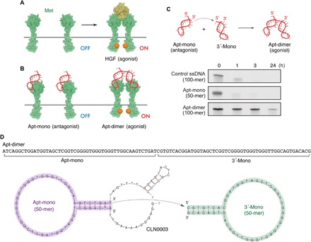
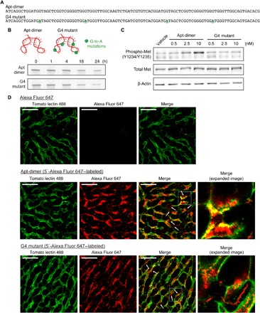
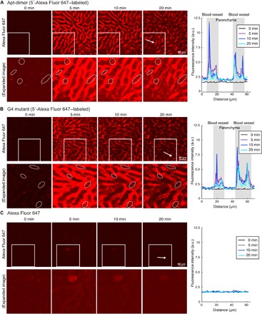
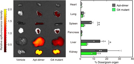
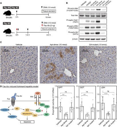
Although growth factors have great therapeutic potential because of their regenerative functions, they often have intrinsic drawbacks, such as low thermal stability and high production cost. Oligonucleotides have recently emerged as promising chemical entities for designing synthetic alternatives to growth factors. However, their applications in vivo have been recognized as a challenge because of their susceptibility to nucleases and limited distribution to a target tissue. Here, we present the first example of oligonucleotide-based growth factor mimetics that exerts therapeutic effects at a target tissue after systemic injection. The aptamer was designed to dimerize a growth factor receptor for its activation and mitigated the progression of Fas-induced fulminant hepatitis in a mouse model. This unprecedented functionality of the aptamer can be reasonably explained by its high nuclease stability and migration to the liver parenchyma. These mechanistic analyses provided insights for the successful application of aptamer-based receptor agonists.


Figures

Fig. 1
Fig. 1. Met-binding DNA aptamers and their nuclease stability.
(A and B) Schematic representation of HGF-induced Met activation and the Met activation potential of the Apt-mono and the Apt-dimer. (C) (Top) Schematic representation of the oligonucleotides used in the present work. (Bottom) Nuclease stability of the oligonucleotides in serum. Each oligonucleotide (2 μM) was incubated in PBS containing 50% FBS at 37°C. (D) Sequence and predicted secondary structure of the aptamers used in the present work. The sequence of the Apt-mono (purple) was identified as the minimal binding motif of a Met-binding aptamer (CLN0003) reported previously (22). The Apt-dimer [termed “ss-0” in a previous work (11)] was designed as a tandem dimer of the Met-binding aptamer, as depicted in the figure. The stem sequence of the 3′-mono (green) was replaced with an alternative complimentary sequence to prevent the misfolding of the aptamer.
Fig. 2
Fig. 2. Design of an unfunctional mutant of the Apt-dimer (G4 mutant) and microdistribution of the oligonucleotides in liver tissues.
(A) Sequences of the oligonucleotides used in the experiments depicted in Fig. 2. The four G-to-A mutations of the G4 mutant are highlighted. (B) Nuclease stability of the Apt-dimer and G4 mutant. Each oligonucleotide (2 μM) was incubated in PBS containing 50% FBS at 37°C. After incubation, the samples were immediately analyzed using denaturing 15% PAGE. (C) Western blotting analysis of the phosphorylation level of Met in SCCVII cells after 15 min of incubation with oligonucleotides. (D) Confocal imaging of liver tissues. Alexa Fluor 647 carboxylic acid (0.5 nmol) or a 5′-Alexa Fluor 647–labeled oligonucleotide (0.5 nmol) was intravenously injected (red). Blood vessels were visualized by injecting 25 μg of DyLight 488–conjugated tomato lectin (green). Ten minutes after the oligonucleotide injection, the liver was excised and observed directly using a confocal laser scanning microscope. Scale bars, 50 μm.
Fig. 3
Fig. 3. Intravital real-time imaging of liver tissues after systemic injection of the oligonucleotides.
(A) 5′-Alexa Fluor 647–labeled Apt-dimer (0.5 nmol), (B) 5′-Alexa Fluor 647–labeled G4 mutant (0.5 nmol), or (C) Alexa Fluor 647 (0.5 nmol) was intravenously injected. (Left) Confocal images. Lower panels show enlarged and brighter images of the boxed region in the upper panels. The dotted circles highlight the leakage of Apt-dimer to the liver parenchyma. (Right) Intensity profile of Alexa Fluor 647 in the region indicated by the white arrows in the left figures. Scale bars, 50 μm. a.u., arbitrary units.
Fig. 4
Fig. 4. Tissue distribution of the aptamer.
Tissue distribution of the 5′-Alexa Fluor 647–labeled oligonucleotides. (Left) Representative ex vivo fluorescence image of the organs 10 min after intravenous injection of Alexa Fluor 647–labeled oligonucleotides (0.5 nmol). The organs were excised after perfusion with PBS and imaged using IVIS (excitation/emission = 640/680 nm). (Right) Biodistribution of oligonucleotides quantified by fluorescence intensity in the supernatant of the organ homogenates. The results are expressed as the mean ± SD (n = 3). Statistical significance was examined by two-sided Student’s t test (**P < 0.01; *P < 0.05).
Fig. 5
Fig. 5. Agonistic functions and therapeutic potential of the HGF-mimic aptamer.
(A) Schematic representation of the experimental outlines. (B) Immunoblotting analysis of Met activation in mouse livers after intravenous injection of recombinant human HGF (rhHGF) or oligonucleotides. The liver was excised 10 min after the intravenous injection of rhHGF (1 μg) or oligonucleotides (1 to 10 nmol). (C) Immunohistochemistry of Met activation in the mouse liver after intravenous injection of the oligonucleotides. The liver was excised 10 min after the intravenous injection of oligonucleotides (10 nmol). (D) (Left) Schematic representation of the apoptotic signal induced by Fas-Ab and the antiapoptotic signal induced by Met activation. (Right) Effect of Apt-dimer administration on a fulminant hepatitis mouse model. An anti–Fas-Ab (2 ng) was co-injected with vehicle control (n = 10), Apt-dimer (10 nmol, n = 9), or G4 mutant (10 nmol, n = 9) intravenously. The oligonucleotide (10 nmol) was administered 1.5 hours later. The average activity of glutamate pyruvate transaminase (GPT), glutamate oxaloacetate transaminase (GOT), and lactate dehydrogenase (LDH) is shown with error bars (SD). Statistical significance was examined by one-way analysis of variance (ANOVA) followed by Tukey’s post hoc test (***P < 0.001; **P < 0.01; ns, not significant).

References

    1. Birchmeier C., Birchmeier W., Gherardi E., Vande Woude G. F., Met, metastasis, motility and more. Nat. Rev. Mol. Cell Biol. 4, 915–925 (2003). - PubMed
    1. Trusolino L., Bertotti A., Comoglio P. M., MET signalling: Principles and functions in development, organ regeneration and cancer. Nat. Rev. Mol. Cell Biol. 11, 834–848 (2010). - PubMed
    1. Nakamura T., Nawa K., Ichihara A., Partial purification and characterization of hepatocyte growth factor from serum of hepatectomized rats. Biochem. Biophys. Res. Commun. 122, 1450–1459 (1984). - PubMed
    1. Michalopoulos G., Houck K. A., Dolan M. L., Luetteke N. C., Control of hepatocyte replication by two serum factors. Cancer Res. 44, 4414–4419 (1984). - PubMed
    1. Michalopoulos G. K., DeFrances M. C., Liver regeneration. Science 276, 60–66 (1997). - PubMed

Publication types

MeSH terms