. 2020 Apr 9;12(1):16.
doi: 10.1186/s13089-020-00163-w.

Quantifying systemic congestion with Point-Of-Care ultrasound: development of the venous excess ultrasound grading system

Affiliations

Quantifying systemic congestion with Point-Of-Care ultrasound: development of the venous excess ultrasound grading system

William Beaubien-Souligny et al. Ultrasound J. .

Abstract

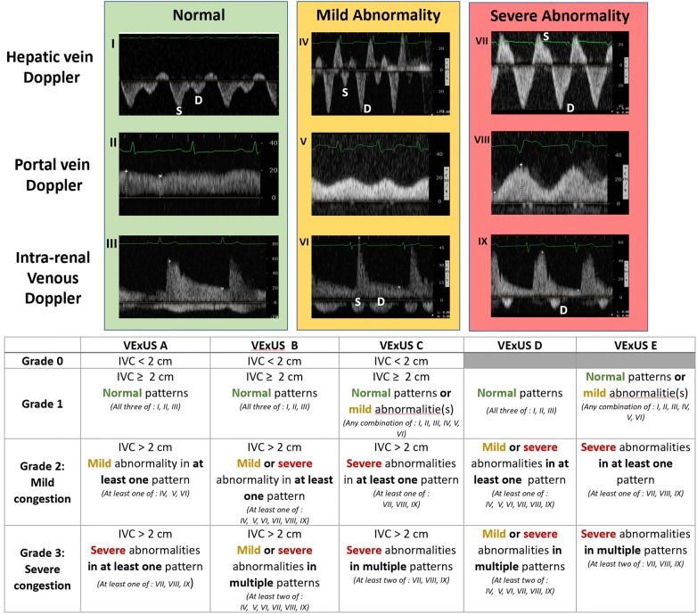
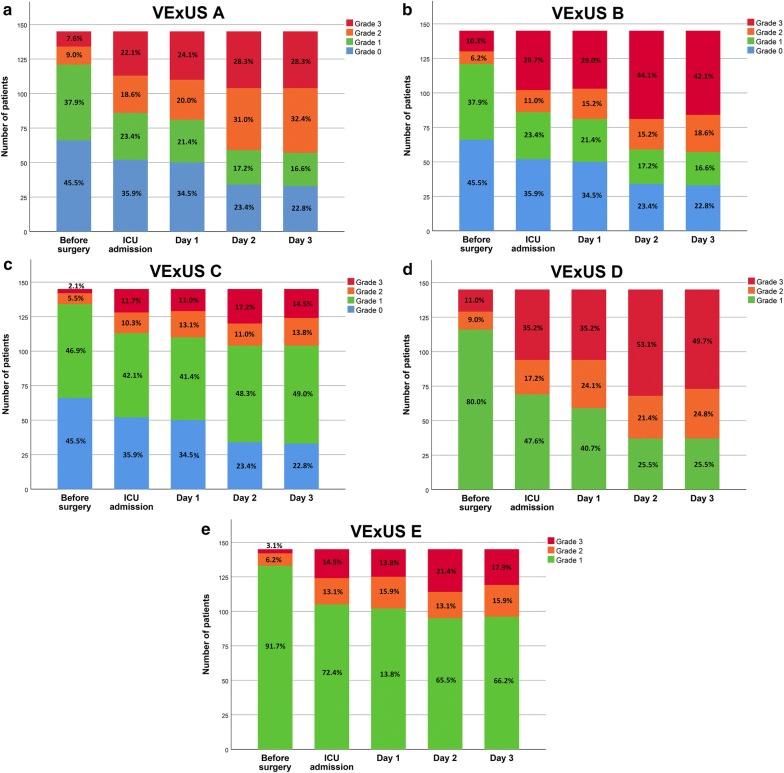
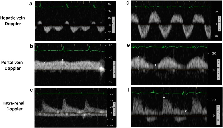
Background: Organ congestion is a mediator of adverse outcomes in critically ill patients. Point-Of-Care ultrasound (POCUS) is widely available and could enable clinicians to detect signs of venous congestion at the bedside. The aim of this study was to develop several grading system prototypes using POCUS and to determine their respective ability to predict acute kidney injury (AKI) after cardiac surgery. This is a post-hoc analysis of a single-center prospective study in 145 patients undergoing cardiac surgery for which repeated daily measurements of hepatic, portal, intra-renal vein Doppler and inferior vena cava (IVC) ultrasound were performed during the first 72 h after surgery. Five prototypes of venous excess ultrasound (VExUS) grading system combining multiple ultrasound markers were developed.

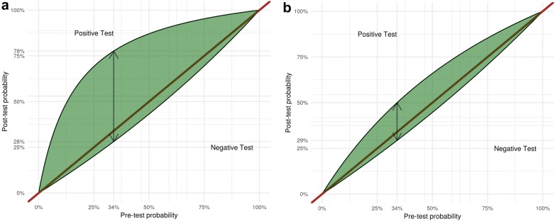
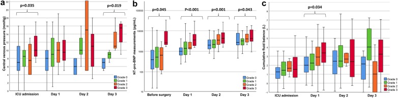
Results: The association between each score and AKI was assessed using time-dependant Cox models as well as conventional performance measures of diagnostic testing. A total of 706 ultrasound assessments were analyzed. We found that defining severe venous congestion as the presence of severe flow abnormalities in multiple Doppler patterns with a dilated IVC (≥ 2 cm) showed the strongest association with the development of subsequent AKI compared with other combinations (HR: 3.69 CI 1.65-8.24 p = 0.001). The association remained significant after adjustment for baseline risk of AKI and vasopressor/inotropic support (HR: 2.82 CI 1.21-6.55 p = 0.02). Furthermore, this severe VExUS grade offered a useful positive likelihood ratio (+LR: 6.37 CI 2.19-18.50) when detected at ICU admission, which outperformed central venous pressure measurements.

Conclusions: The combination of multiple POCUS markers may identify clinically significant venous congestion.

Keywords: Acute kidney injury; Cardiac surgery; Fluid balance; Point-Of-Care ultrasound; Venous congestion.


Conflict of interest statement

André Y. Denault is a Speaker for CAE Healthcare, Edwards and Masimo. The other authors have no conflict of interest to declare.

Figures

Fig. 1
Fig. 1
The Venous Excess UltraSound (VExUS) grading system prototypes combining inferior vena cava (IVC) diameter and venous Doppler waveform of the portal, hepatic and interlobular renal veins. Hepatic Doppler is considered mildly abnormal when the systolic (S) component is lower in magnitude than the diastolic (D) component, but still toward the liver while it is considered severely abnormal when the S component is reversed (toward the heart). Portal Doppler is considered mildly abnormal when a variation in the velocities during the cardiac cycle of 30 to < 50% are observed, while is considered severely abnormal when a variation of ≥ 50% is seen. Intra-renal venous Doppler is considered mildly abnormal when it is discontinuous with a systolic (S) and diastolic (D) phase, while is it considered severely abnormal when it is discontinuous with only a diastolic phase seen during the cardiac cycle
Fig. 2
Fig. 2
Distribution of Venous Excess UltraSound (VExUS) grading system prototypes a, b, c, d and e in the perioperative period in 145 patients undergoing cardiac surgery
Fig. 3
Fig. 3
Leaf plots displaying the relationship between the assumed pre-test probability (on the x-axis) and the post-test probability (on the y-axis) of acute kidney injury (AKI) for the following cut-off: a severe congestion (Grade 3) defined by the VExUS C grading system and b central venous pressure of ≥ 12 mmHg. The upper half part of the curve indicates the post-test probability in case of a positive result while the lower half indicated is for a negative test result. The dashed double-sided arrow indicated the test performance considering the incidence of acute kidney injury (pre-rest probability) within the studied cohort (33.8%)
Fig. 4
Fig. 4
Clinical parameters in relationship with the Venous Excess UltraSound (VExUS) grading system C during the peri-operative period. a Central venous pressure at the time of ultrasound assessment in relationship with VExUS C grading system. b N-terminal pro-beta natriuretic peptide (NT-pro-BNP) in relationship with VExUS C grading system. c Cumulative fluid balance in relationship with VExUS C grading system. Significant results (p < 0.05) are highlighted. Complete results of comparisons are presented in Additional file 1: Table S3
Fig. 5
Fig. 5
Example of VExUS C grading system assessment in cardiac surgery. Patient #1: A 55-year-old woman undergoing tricuspid valve repair aortic valve replacement and mitral valve replacement known with chronic kidney disease (baseline eGFR = 35 mL/min/1.73 m2) with left ventricular ejection fraction of 40% and a high risk of major complications (EuroSCORE II = 16.8%) presented the following ultrasound findings at ICU admission after surgery: a Normal hepatic triphasic pattern, b a non-pulsatile portal flow and c continuous intral-renal venous flow and an IVC diameter of > 2.1 cm (not shown) corresponding to Grade 1 of VExUS C grading system. The patient did not develop acute kidney injury, was extubated 2.2 h after ICU admission and was discharged from the ICU less than 24 h after surgery. Patient #2: A 70-year-old man undergoing mitral valve repair with a left ventricular ejection fraction of 50% and a moderate risk of major complication (EuroSCORE II = 1.54%) presented the following ultrasound findings at ICU admission after surgery complicated by right ventricular dysfunction after cardiopulmonary bypass: d Systolic reversal of the hepatic venous flow, e severe portal flow pulsatility and f severe alteration in intra-renal venous flow corresponding to Grade 3 of the VExUS C grading system. The patient developed severe acute kidney injury and delirium in the post-operative period

References

    1. Seymour CW, Rosengart MR. Septic Shock: advances in diagnosis and treatment. JAMA. 2015;314:708–717. doi: 10.1001/jama.2015.7885. - DOI - PMC - PubMed
    1. van Diepen S, Katz Jason N, Albert Nancy M, Henry Timothy D, Jacobs Alice K, Kapur Navin K, Kilic A, Menon V, Ohman EM, Sweitzer Nancy K, Thiele H, Washam Jeffrey B, Cohen Mauricio G. Contemporary management of cardiogenic shock: a scientific statement from the American Heart Association. Circulation. 2017;136:e232–e268. doi: 10.1161/CIRCULATIONAHA.117.029532. - DOI - PubMed
    1. Squara P, Hollenberg S, Payen D. Reconsidering vasopressors for cardiogenic shock: everything should be made as simple as possible, but not simpler. Chest. 2019;156:392–401. doi: 10.1016/j.chest.2019.03.020. - DOI - PubMed
    1. De Backer D, Orbegozo Cortes D, Donadello K, Vincent J-L. Pathophysiology of microcirculatory dysfunction and the pathogenesis of septic shock. Virulence. 2014;5:73–79. doi: 10.4161/viru.26482. - DOI - PMC - PubMed
    1. Saito S, Uchino S, Takinami M, Uezono S, Bellomo R. Postoperative blood pressure deficit and acute kidney injury progression in vasopressor-dependent cardiovascular surgery patients. Crit Care. 2016;20:74. doi: 10.1186/s13054-016-1253-1. - DOI - PMC - PubMed

LinkOut - more resources