. 2020 Mar:15:241-257.
doi: 10.2217/fmb-2019-0165.

Virulence of the emerging pathogen, Burkholderia pseudomallei, depends upon the O-linked oligosaccharyltransferase, PglL

Affiliations

Virulence of the emerging pathogen, Burkholderia pseudomallei, depends upon the O-linked oligosaccharyltransferase, PglL

Samuel J Willcocks et al. Future Microbiol. 2020 Mar.

Abstract

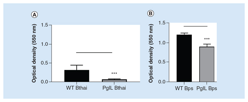
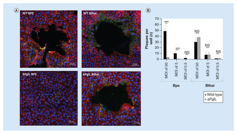
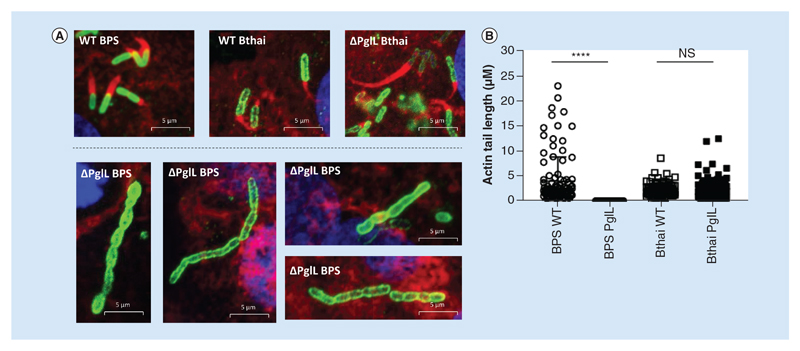
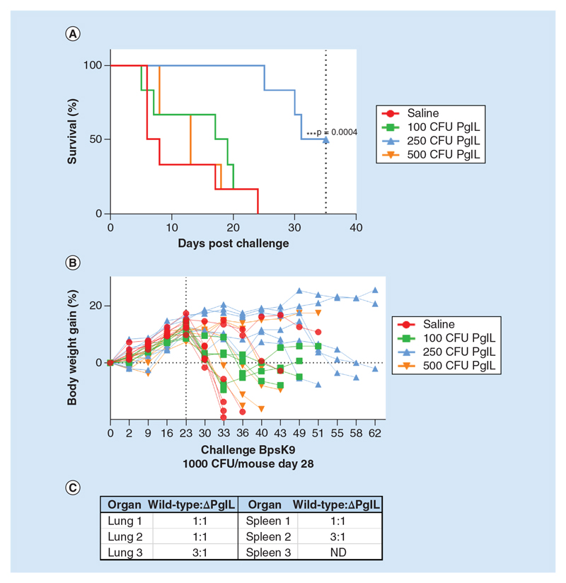
Aim: We sought to characterize the contribution of the O-OTase, PglL, to virulence in two Burkholderia spp. by comparing isogenic mutants in Burkholderia pseudomallei with the related species, Burkholderia thailandensis. Materials & methods: We utilized an array of in vitro assays in addition to Galleria mellonella and murine in vivo models to assess virulence of the mutant and wild-type strains in each Burkholderia species. Results: We found that pglL contributes to biofilm and twitching motility in both species. PglL uniquely affected morphology; cell invasion; intracellular motility; plaque formation and intergenus competition in B. pseudomallei. This mutant was attenuated in the murine model, and extended survival in a vaccine-challenge experiment. Conclusion: Our data support a broad role for pglL in bacterial fitness and virulence, particularly in B. pseudomallei.

Keywords: Burkholderia; PglL; glycosylation; oligosaccharyltransferase; secretion system; virulence.


Conflict of interest statement

Financial & competing interests disclosure

We thank the Wellcome Trust, UK for funding this research (grant ref 102979/Z/13/Z). The authors have no other relevant affiliations or financial involvement with any organization or entity with a financial interest in or financial conflict with the subject matter or materials discussed in the manuscript apart from those disclosed.

No writing assistance was utilized in the production of this manuscript.

Figures

Figure 1
Figure 1. Biofilm formation in wild-type and ΔpglL Burkholderia spp. B.
thailandensis E264 (A) or B. pseudomallei K9264 (B) strains were incubated statically in peg-assay 96-well plates as described at 37°C for 48 h. Biofilm formation was assessed by crystal violet staining and measurement of optical density at 550 nm. Student’s t-test was performed for each species’ wild-type versus the mutant strain using GraphPad Prism 8.1.2. Error bars represent standard deviation from the mean of seven technical repeats. A representative figure from three independent biological replicates is shown for each species. ***p < 0.001.
Figure 2
Figure 2. Twitching motility of wild-type and ΔpglL Burkholderia spp. B.
pseudomallei K9264 (A) or B. thailandensis E264 (B) strains were grown between a plastic-agarose interface at room temperature and resultant mean colony circumference measured. Student’s t-test was performed for each species’ wild-type versus the mutant strain using GraphPad Prism 8.1.2. Error bars represent standard deviation from the mean of triplicate technical repeats. A representative figure from three independent biological replicates is shown. ***p < 0.001.
Figure 3
Figure 3. Sensitivity of wild-type and ΔpglL Burkholderia spp. to Polymixin B and serum. B.
thailandensis E264 or B. pseudomallei K9264 strains were incubated with a titration of either human serum (A & B); heat-treated serum (C & D); or polymixin B (E & F). Alternatively, human serum was heat inactivated by treatment at 65°C for 1 h. Student’s t-test was performed for each species’ wild-type versus the mutant strain within each condition using GraphPad Prism 8.1.2. Error bars represent standard deviation from the mean of triplicate technical repeats. A representative figure from three independent biological replicates is shown. *p < 0.05; **p < 0.01; ***p < 0.001. CFU: Colony-forming unit.
Figure 4
Figure 4. Attachment, uptake and intracellular survival of wild-type and ΔpglL Burkholderia spp.
Burkholderia spp. strains were used to infect either human epithelial A549 cells or murine Raw 264.7 cells at an multiplicity of infection of five for 90 min before washing, lysis and enumeration of bacteria by colony-forming unit assay (A). Raw 264.7 cells were infected for longer time periods as described, with kanamycin used to control extracellular bacterial replication. Values are expressed as proportion of cells versus the infective dose (A) or versus 90 min colony-forming unit (B). Student’s ŕ-test of wild-type versus mutant within each group was performed using GraphPad Prism 8.1.2. Error bars represent standard deviation from the mean of triplicate technical repeats. A representative figure from three independent biological replicates is shown. **p < 0.05; ***p < 0.001. WT: Wild-type.
Figure 5
Figure 5. Plaque formation by wild-type and ΔpglL Burkholderia spp. In an A549 cell monolayer.
Burkholderia spp. strains were used to infect a confluent monolayer of A549 cells in chamber slides. After 90 min infection at different multiplicity of infections, cells were incubated in the presence of 100 μg ml-1 kanamycin for a further 16 h. Immunofluorescence with confocal microscopy was used to image the monolayers, with nuclei stained with DAPI (blue), host-cell actin cytoskeleton was stained with phalloidin-alexafluor-546 (red) and bacteria visualized using an alexafluor-488 secondary antibody (A). For counting, plaques were visualized using phase-contrast microscopy and the entire well of duplicate wells was used for counting for each condition (B). Student’s t-test of wild-type versus mutant was performed within each species and multiplicity of infection group using GraphPad Prism 8.1.2. Error bars represent standard deviation from the mean of duplicate technical repeats. A representative figure from two independent biological replicates is shown. ***p < 0.001. WT: Wild-type.
Figure 6
Figure 6. Actin-tail formation by wild-type and ΔpglL Burkholderia spp. in an A549 cell monolayer.
Burkholderia spp. strains were used to infect a confluent monolayer of A549 cells in chamber slides. After 90 min infection at different multiplicity of infections, cells were incubated in the presence of 100 μg ml-1 kanamycin for a further 16 h. Immunofluorescence with confocal microscopy was used to image the monolayers, with nuclei stained with DAPI (blue), host-cell actin cytoskeleton was stained with phalloidin-alexafluor-546 (red) and bacteria visualized using an alexafluor-488 secondary antibody (A). To measure actin tails, Zeiss Zen software was used, and the results displayed as a scatter chart (B). Mean lengths were: BPS WT 3916 nm (n = 106); BPS ΔpglL 3 nm (n = 43); Bthai WT 2204 nm (n = 86); Bthai ΔpglL 2222 nm (n = 112). Statistical difference between mutant and wild-type for each species was determined by Mann–Whitney U test using GraphPad Prism 8.1.2. ****p < 0.0001.
Figure 7
Figure 7. Competition assay between wild-type or ΔpglL Burkholderia spp. and E. coli.
Burkholderia spp. strains were cocultured with E. coli_pcDNA 3.3 TOPO_LacZ at identical optical densities for 5 h in either LB broth or spotted onto LB agar, as indicated. Bacteria were enumerated by colony-forming unit assay on agar plates coated with X-gal for distinguishing bacterial species by blue/white screening. Student’s t-test was performed for each species’ wild-type versus the mutant strain within each condition. Using GraphPad Prism 8.1.2. Error bars represent standard deviation from the mean of triplicate technical repeats. A representative figure from three independent biological replicates is shown. *p < 0.05; ***p < 0.001. CFU: Colony-forming unit; WT: Wild-type.
Figure 8
Figure 8. In vivo virulence of wild-type (WT) and ΔpglL Burkholderia spp.
G. mellonella larvae (n = 10 per group) were infected with 1000 colony-forming unit Bthai WT or Bthai ΔpglL or inoculated with PBS alone. After 36 h, difference between WT E264 survival was compared with mutant pglL (A). Female BALB/C mice (n = 5 per group) were infected intranasally with approximately 1000 colony-forming unit either WT BPS K92643 or BPS ΔpglL in sterile saline (B). Surviving mice were culled at day 30 post infection and total bacterial colony-forming unit in lungs and spleen enumerated (C). **p < 0.05 Log-rank Mantel-Cox and Gehan-Breslow-Wilcoxon test; ***p < 0.01 Log-rank Mantel-Cox and Gehan-Breslow-Wilcoxon test. CFU: Colony-forming unit; WT: Wild-type.
Figure 9
Figure 9. Vaccination of female BALB/C mice with ΔpglL B. pseudomallei and challenge with wild-type BPS K9264.
Female BALB/C mice (n = 6 per group) were infected intranasally with different colony-forming unit of BPS ΔpglL as indicated, and after 28 days, challenged intranasally with wild-type BPS K9264. Survival (A) and body weight (B) were monitored up to 68 days post vaccination. At 68 days, surviving mice (from the 250 CFU vaccinated group) were culled and strains of organ-resident bacteria identified by polymerase chain reaction for the pglL gene to establish the ratio of wild-type (challenge strain) to ΔpglL (vaccine strain) colonies (C). ***p < 0.01 Log-rank Mantel-Cox and Gehan-Breslow-Wilcoxon test. CFU: Colony-forming unit; ND: Excluded due to splenomegaly.

References

    1. Angus AA, Agapakis CM, Fong S, et al. Plant-associated symbiotic Burkholderia species lack hallmark strategies required in mammalian pathogenesis. PLoS ONE. 2014;9(1):e83779. - PMC - PubMed
    1. Jilani MS, Robayet JA, Mohiuddin M, Hasan MR, Ahsan CR, Haq JA. Burkholderia pseudomallei its detection in soil and seroprevalence in Bangladesh. PLoS Negl Trop Dis. 2016;10(1):e0004301. - PMC - PubMed
    1. Steinmetz I, Wagner GE, Kanyala E, et al. Melioidosis in Africa: time to uncover the true disease load. Trop Med Infect Dis. 2018;3(2):pii:E62 - PMC - PubMed
    1. Wuthiekanun V, Mayxay M, Chierakul W, et al. Detection of Burkholderia pseudomallei in soil within the Lao People’s Democratic Republic. J Clin Microbiol. 2005;43(2):923–924. - PMC - PubMed
    1. Rolim DB, Vilar DC, Sousa AQ, et al. Melioidosis, northeastern Brazil. Emerg Infect Dis. 2005;11(9):1458–1460. - PMC - PubMed

Publication types

Substances

LinkOut - more resources