. 2020 Apr 9;5(7):e134564.
doi: 10.1172/jci.insight.134564.

Pancreatic tropism of metastatic renal cell carcinoma

Affiliations

Pancreatic tropism of metastatic renal cell carcinoma

Nirmish Singla et al. JCI Insight. .

Abstract

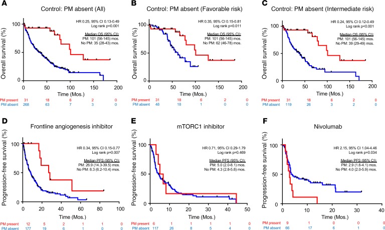
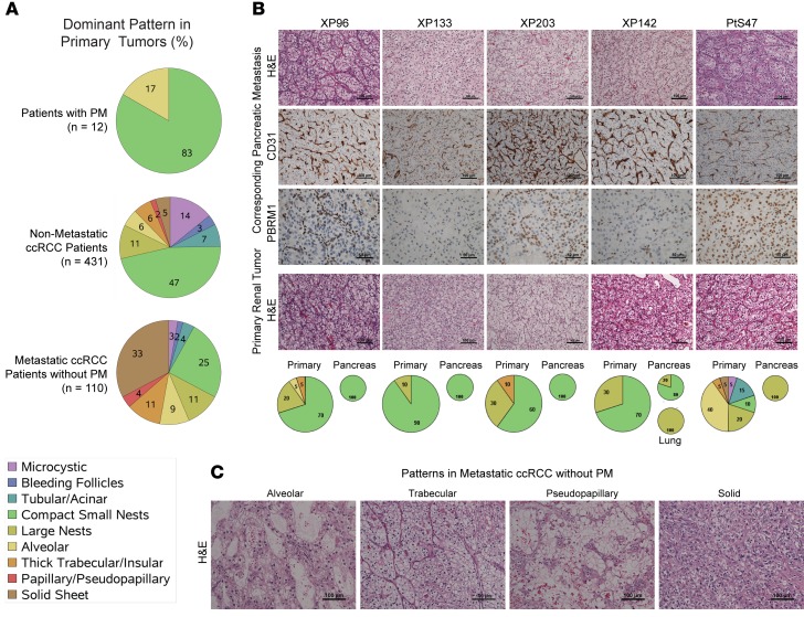
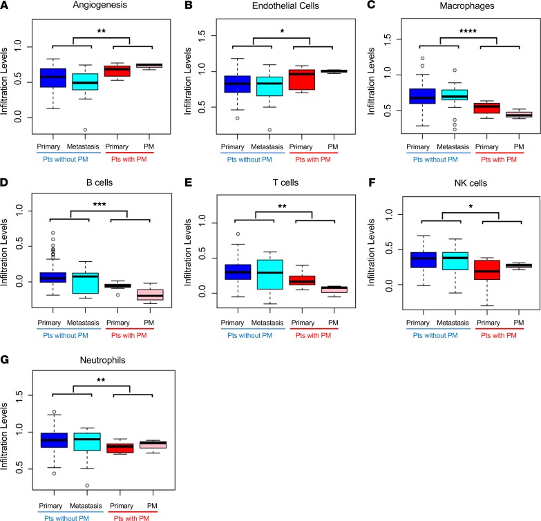
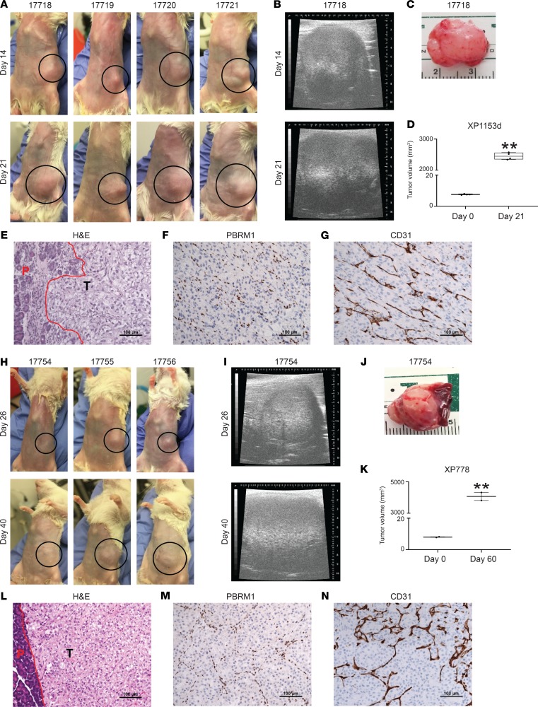
Renal cell carcinoma (RCC) is characterized by a particularly broad metastatic swath, and, enigmatically, when the pancreas is a destination, the disease is associated with improved survival. Intrigued by this observation, we sought to characterize the clinical behavior, therapeutic implications, and underlying biology. While pancreatic metastases (PM) are infrequent, we identified 31 patients across 2 institutional cohorts and show that improved survival is independent of established prognostic variables, that these tumors are exquisitely sensitive to antiangiogenic agents and resistant to immune checkpoint inhibitors (ICIs), and that they are characterized by a distinctive biology. Primary tumors of patients with PM exhibited frequent PBRM1 mutations, 3p loss, and 5q amplification, along with a lower frequency of aggressive features such as BAP1 mutations and loss of 9p, 14q, and 4q. Gene expression analyses revealed constrained evolution with remarkable uniformity, reduced effector T cell gene signatures, and increased angiogenesis. Similar findings were observed histopathologically. Thus, RCC metastatic to the pancreas is characterized by indolent biology, heightened angiogenesis, and an uninflamed stroma, likely underlying its good prognosis, sensitivity to antiangiogenic therapies, and refractoriness to ICI. These data suggest that metastatic organotropism may be an indicator of a particular biology with prognostic and treatment implications for patients.

Keywords: Oncology; Urology.


Conflict of interest statement

Conflict of interest: JB is a consultant for Exelixis and received research funding from Genentech, Peloton, and Arrowhead. IP is a consultant for Bayer Healthcare. BR serves as a consultant for and received research funding from Pfizer, Merck, GNE/Roche, Peloton, Aveo, and BMS. BR also received research funding from AstraZeneca and serves as a consultant for Novartis, Synthorx, Compugen, Corvus, and Exelixis. BR owns stock in PTC Therapeutics.

Figures

Figure 1
Figure 1. Patients with PM have improved survival that is independent of the IMDC risk score and better disease control with angiogenesis inhibitors compared with other treatments.
(A) Kaplan-Meier survival analyses of PM cohort compared with a historical control of 268 metastatic ccRCC without PM. Kaplan-Meier survival analyses of PM cohort compared with a historical control in (B) favorable (n = 48) or (C) intermediate (n = 119) IMDC risk groups. Time is measured from metastatic diagnosis. (D) PFS in metastatic ccRCC patients treated with first-line angiogenic inhibitors, stratified by the presence (n = 12) or absence (n = 177) of PM. PFS with (E) mTORC1 inhibitors (6 patients with vs. 117 patients without PM) and (F) nivolumab (9 patients with vs. 66 patients without PM). PM, pancreatic metastases; ccRCC, clear cell renal cell carcinoma; IMDC, International Metastatic Database Consortium; PFS, progression-free survival; mTORC1, mTOR complex 1.
Figure 2
Figure 2. PM tumors are characterized by a homogeneous, small nest architectural pattern with a highly vascular network and infrequent inflammatory cells.
(A) Pie charts showing the distribution of architectures in primary tumors of patients with PM (n = 12) and reference pathology cohorts: patients without metastases (n = 431), and patients with non-PM metastases (n = 110). (B) H&E sections and IHC for PBRM1 and CD31 of PM as well as H&E sections of corresponding primary tumors. Pie charts of architectures corresponding to patients are shown below. (C) Architectural subtypes associated with more aggressive ccRCC. PM, pancreatic metastases; ccRCC, clear cell renal cell carcinoma.
Figure 3
Figure 3. PM tumors are characterized by a mutational and copy number profile of less aggressive ccRCC, with clustered gene expression revealing constrained evolution.
(A) Oncoplot of somatic mutations based on the Catalogue of Somatic Mutations in Cancer in primary tumors (T suffix) and metastases (M suffix) from 31 patients with PM (separated by white lines). Most highly mutated genes are shown, with corresponding patient mutation percentages (left). (B) Average copy number alterations of primary and metastatic samples (n = 48) corresponding to 31 patients. (C) Principal component analysis of gene expression from fresh frozen PM primary tumors and metastases (n = 12) compared with controls (UT Southwestern) (Hotelling’s T-squared P < 1 × 10–16). PM, pancreatic metastases; ccRCC, clear cell renal cell carcinoma.
Figure 4
Figure 4. Primary tumors with PM are characterized by an angiogenic signature and low levels of inflammation.
Box-and-whisker plots comparing median relative gene expression levels (lines within the boxes) and IQRs (bounds of the boxes) for primary tumors and metastases of PM and non-PM patients for the indicated empirically defined tumor microenvironment gene signatures. Whiskers extend to 1.5 times the IQR. Open circles represent the most extreme data points beyond the whiskers (*P < 0.05, **P < 0.01, ***P < 0.001, ****P < 0.0001 by 2-tailed Student’s t tests). PM, pancreatic metastases; IQR, interquartile range.
Figure 5
Figure 5. The pancreas supports engraftment and growth of ccRCC with PBRM1 loss.
(A and H) Images of tumors growing in the pancreas of 7 NOD/SCID host mice from 2 different patients with PBRM1 mutations. Dotted black circles demarcate the tumor areas. (B and I) Ultrasound images of tumor growth. (C and J) Images of harvested tumors from indicated mice. (D and K) Tumor volumes at implantation and harvest. (E and L) H&E staining of the tumors showing pancreatic interface in red; P-pancreas; T-tumor. (F and M) PBRM1 and (G and N) CD31 IHC studies. **P < 0.01.

References

    1. Villarreal-Garza C, Perez-Alvarez SI, Gonzalez-Espinoza IR, Leon-Rodriguez E. Unusual metastases in renal cell carcinoma: a single institution experience and review of literature. World J Oncol. 2010;1(4):149–157. - PMC - PubMed
    1. Sountoulides P, Metaxa L, Cindolo L. Atypical presentations and rare metastatic sites of renal cell carcinoma: a review of case reports. J Med Case Rep. 2011;5:429. - PMC - PubMed
    1. Bianchi M, et al. Distribution of metastatic sites in renal cell carcinoma: a population-based analysis. Ann Oncol. 2012;23(4):973–980. doi: 10.1093/annonc/mdr362. - DOI - PubMed
    1. Sperti C, et al. Metastatic tumors to the pancreas: a systematic review and meta-analysis. Minerva Chir. 2016;71(5):337–344. - PubMed
    1. Sperti C, Moletta L, Patanè G. Metastatic tumors to the pancreas: The role of surgery. World J Gastrointest Oncol. 2014;6(10):381–392. doi: 10.4251/wjgo.v6.i10.381. - DOI - PMC - PubMed

Publication types

MeSH terms