Review
. 2020 Apr 10;126(8):1086-1106.
doi: 10.1161/CIRCRESAHA.119.315862. Epub 2020 Apr 9.

Cardiomyocyte Maturation: New Phase in Development

Affiliations
Review

Cardiomyocyte Maturation: New Phase in Development

Yuxuan Guo et al. Circ Res. .

Abstract

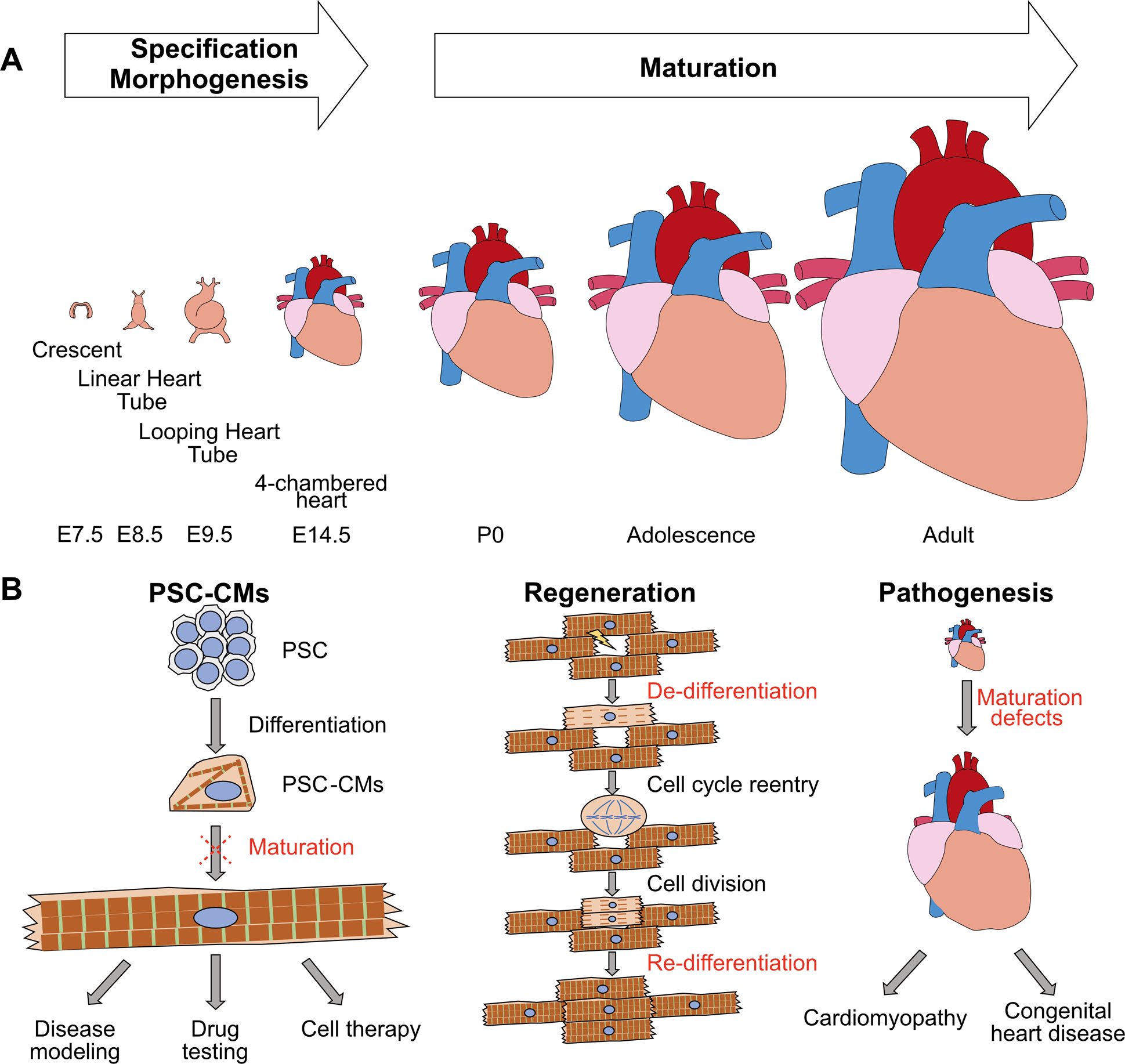
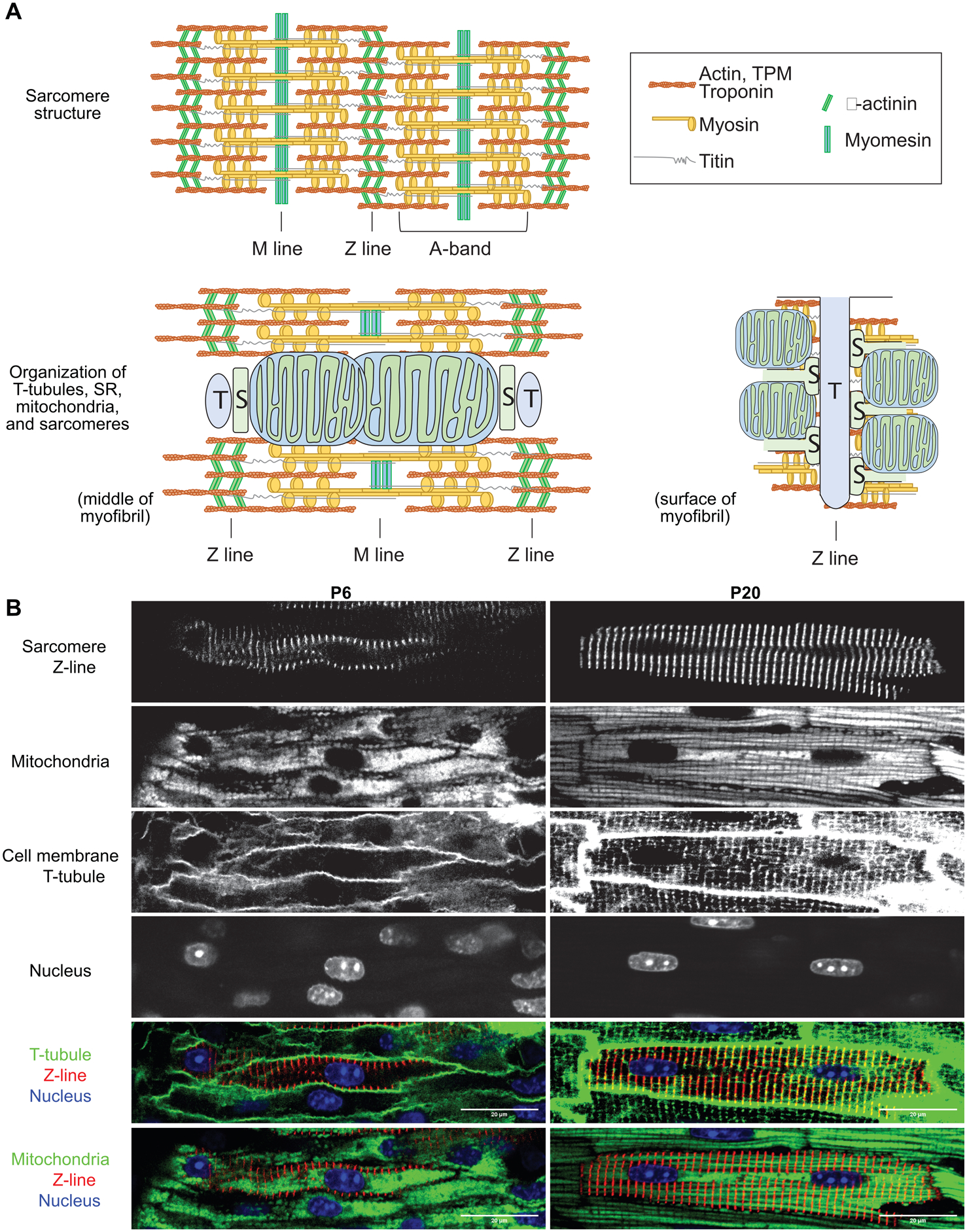
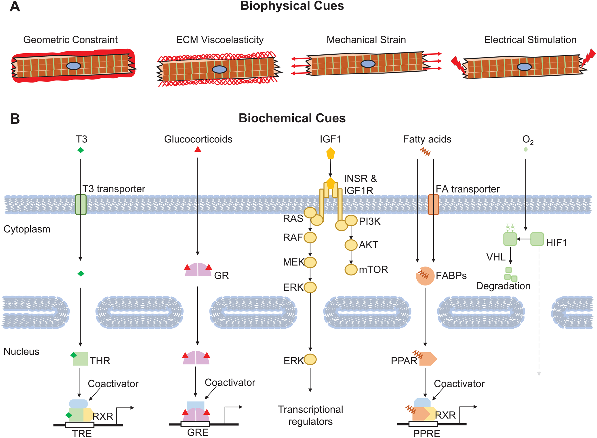
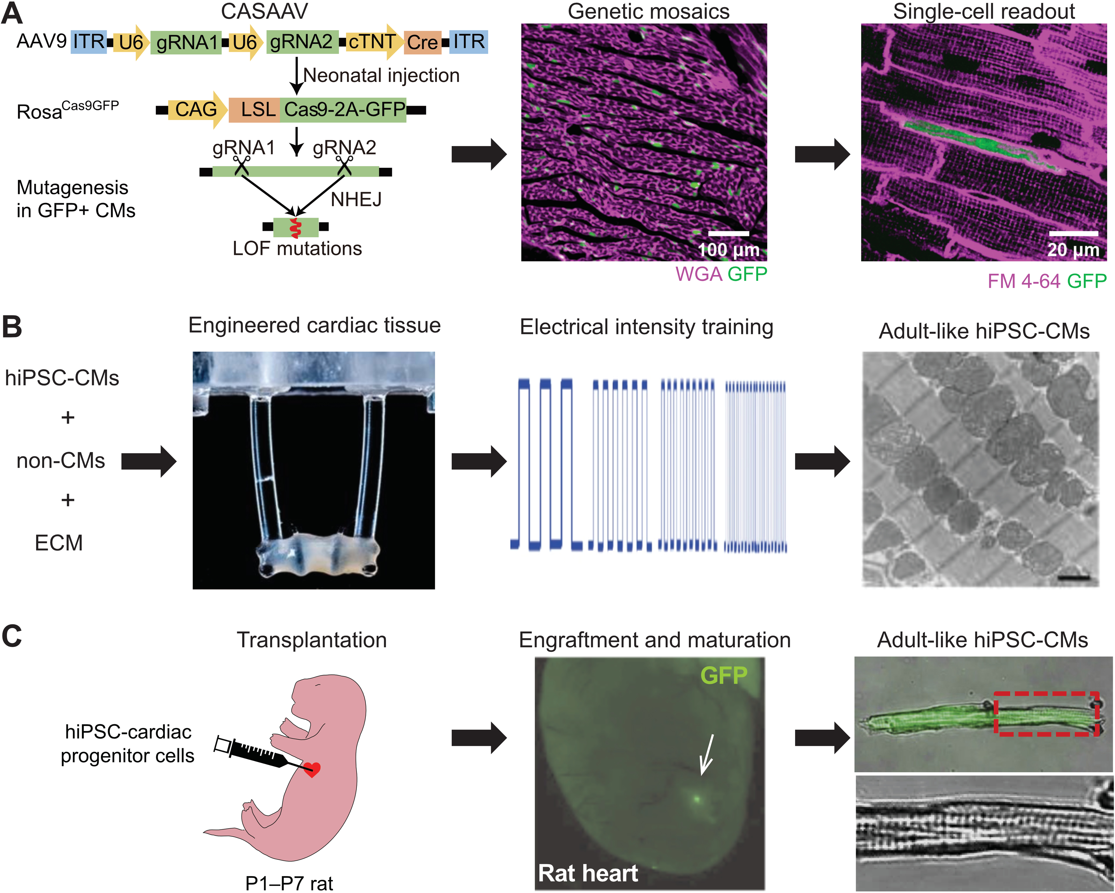
Maturation is the last phase of heart development that prepares the organ for strong, efficient, and persistent pumping throughout the mammal's lifespan. This process is characterized by structural, gene expression, metabolic, and functional specializations in cardiomyocytes as the heart transits from fetal to adult states. Cardiomyocyte maturation gained increased attention recently due to the maturation defects in pluripotent stem cell-derived cardiomyocyte, its antagonistic effect on myocardial regeneration, and its potential contribution to cardiac disease. Here, we review the major hallmarks of ventricular cardiomyocyte maturation and summarize key regulatory mechanisms that promote and coordinate these cellular events. With advances in the technical platforms used for cardiomyocyte maturation research, we expect significant progress in the future that will deepen our understanding of this process and lead to better maturation of pluripotent stem cell-derived cardiomyocyte and novel therapeutic strategies for heart disease.

Keywords: heart disease; mammals; pluripotent stem cell; regeneration; stem cell.


Figures

Figure 1.
Figure 1.. Heart maturation and its implication in translational medicine.
(A) Conceptual scheme of the maturation phase of heart development. Mouse stages are labeled at bottom. (B) Major applications of CM maturation studies. Left: to promote the maturation of PSC-CMs. Mid: to optimize CM regeneration conditions. Right: to better understand cardiac pathogenesis.
Figure 2.
Figure 2.. Structural maturation of CMs.
(A) A schematic view of sarcomere components in mature CMs (top) and spatial relationship between sarcomeres and T-tubule (T), SR (S) and mitochondria in mature CMs (bottom). Bottom left: a view across the middle of a myofibril. Bottom right: a view on the cytoplasmic surface of a myofibril. (B) In situ confocal images of murine myocardium at postnatal day 6 (P6) and P20. Sarcomere Z-lines were labeled by AAV-Actn2-GFP infection. Mitochondria, T-tubules and nuclei were stained by TMRM (polarized mitochondria), FM 4–64 (plasma membrane), and Hoechst (DNA), respectively, through Langendoff perfusion. Merged images highlight T-tubule-sarcomere and mitochondria-sarcomere associations that are established during postnatal maturation.
Figure 3.
Figure 3.. Representative environmental cues that regulate CM maturation.
(A) Key biophysical factors that affect CM maturation. (B) Critical biochemical cues that regulate CM maturation. Representative signal receptors, messengers and transcriptional regulators are also depicted.
Figure 4.
Figure 4.. Model systems to study CM maturation.
(A) CASAAV-based genetic mosaic analysis of murine CM maturation in vivo. Expression of genome-encoded Cas9-P2A-GFP was activated by AAV-delivery of single or dual gRNAs and Cre, expressed from the cardiomyocyte-specific cTNT promoter (left). When the AAV is given at a low dose, mosaic transduction and Cas9-mediated somatic mutagenesis at genes targeted by gRNA(s) occurs (GFP+ cells, middle). The phenotype of single GFP+ cells is then analyzed (right, illustrating T-tubule and maturational growth defects caused by Srf depletion). WGA, wheat germ agglutinin. FM 4–64, membrane dye. Left panel reprinted ref. 21. with permission. (B) In vitro maturation of PSC-CMs by tissue engineering and electrical pacing. 3D cultured engineered heart tissue was assembled from PSC-CMs (left). Elastomeric posts apply anisotropic stress on muscle bundle. Rapid electrical pacing protocol was applied from early in the PSC-CM differentiation process (middle), resulting in well organized, mature PSC-CMs, as evaluated by transmission electron microscopy (right). Reprinted from ref. 4 with permission. (C) In vivo maturation of PSC-CMs. Human PSC-CMs expressing GFP were injected into the hearts of immunodeficient neonatal rats (left). After several weeks, engrafted PSC-CMs (GFP+, middle) have mature morphology (right). Reprinted from ref. with permission.

References

    1. Tu C, Chao BS and Wu JC. Strategies for Improving the Maturity of Human Induced Pluripotent Stem Cell-Derived Cardiomyocytes. Circ Res. 2018;123:512–514. - PMC - PubMed
    1. Kannan S and Kwon C. Regulation of cardiomyocyte maturation during critical perinatal window. J Physiol. 2018. - PMC - PubMed
    1. Huang CY, Maia-Joca RPM, Ong CS, Wilson I, DiSilvestre D, Tomaselli GF and Reich DH. Enhancement of human iPSC-derived cardiomyocyte maturation by chemical conditioning in a 3D environment. Journal of Molecular and Cellular Cardiology. 2019. - PubMed
    1. Ronaldson-Bouchard K, Ma SP, Yeager K, Chen T, Song L, Sirabella D, Morikawa K, Teles D, Yazawa M and Vunjak-Novakovic G. Advanced maturation of human cardiac tissue grown from pluripotent stem cells. Nature. 2018;556:239–243. - PMC - PubMed
    1. Karbassi E, Fenix A, Marchiano S, Muraoka N, Nakamura K, Yang X and Murry CE. Cardiomyocyte maturation: advances in knowledge and implications for regenerative medicine. Nat Rev Cardiol. 2020:10.1038/s41569-019-0331-x. - DOI - PMC - PubMed

Publication types