Review
. 2020 Apr 10;126(8):1061-1085.
doi: 10.1161/CIRCRESAHA.120.315895. Epub 2020 Apr 9.

Hematopoiesis and Cardiovascular Disease

Affiliations
Review

Hematopoiesis and Cardiovascular Disease

Wolfram C Poller et al. Circ Res. .

Abstract

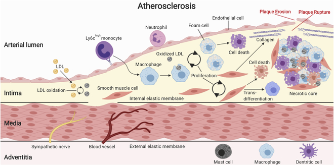
A central feature of atherosclerosis, the most prevalent chronic vascular disease and root cause of myocardial infarction and stroke, is leukocyte accumulation in the arterial wall. These crucial immune cells are produced in specialized niches in the bone marrow, where a complex cell network orchestrates their production and release. A growing body of clinical studies has documented a correlation between leukocyte numbers and cardiovascular disease risk. Understanding how leukocytes are produced and how they contribute to atherosclerosis and its complications is, therefore, critical to understanding and treating the disease. In this review, we focus on the key cells and products that regulate hematopoiesis under homeostatic conditions, during atherosclerosis and after myocardial infarction.

Keywords: atherosclerosis; hematopoiesis; leukocytes; myocardial infarction; stem cell.


Figures

Figure 1:
Figure 1:. The hematopoietic stem cell niche.
HSCs mainly reside in the bone marrow, where the bulk of hematopoiesis occurs during adulthood. Bone structures that form and protect the medullary cavity are densely innervated and vascularized with the periosteum lining the outer surface and the endosteum coating the inner surface. The endosteum forms the interface between bone and bone marrow and consists of bone-lining cells including osteoblasts that form bone substance and osteoclasts that resorb it. Arteries reach the bone marrow via nutrient canals, branch into smaller arteries and eventually into thin-walled arterioles. Transitional vessels connect arterioles with an extensive sinusoidal network that drains into a central venous sinus, which in turn empties into nutrient veins that leave the marrow via nutrient canals. Sympathetic fibers follow the arteries into the bone marrow, where they form a structural network with neural-glial antigen- (NG2) and nestin- (Nes) positive perivascular stromal cells. HSCs primarily reside in perivascular niches around fenestrated sinusoids as well as around less-permeable arterioles, while lymphoid progenitors reside in an osteoblastic niche closer to the endosteum. Overall, the bone marrow niche is formed by a complex network of non-hematopoietic as well as hematopoietic cell types that produce multiple factors orchestrating maintenance, proliferation and differentiation of HSCs and their progeny. The best-studied and apparently most relevant niche cell types are endothelial cells (sinusoidal and arteriolar ECs) and perivascular mesenchymal stromal cells (Ng2-CreER+, LEPR+/CAR cells) that produce crucial HSC maintenance factors such as SCF and CXCL12. Other cell types involved in niche formation include osteoblasts, osteoclasts, sympathetic nerve fibers, non-myelinating Schwann cells, adipocytes, megakaryocytes, macrophages and regulatory T cells (Treg). Abbreviations: NG2 - neuron/glial antigen 2; HSC - hematopoietic stem cell; Treg - regulatory T cell; LEPR - leptin receptor; CAR cell - CXCL12-abundant reticular cell; EC - endothelial cell; CXCL12 - CXC chemokine ligand 12; SCF - stem cell factor; OPN - osteopontin; IL7 - interleukin-7; TGFβ - transforming growth factor-β; CXCL4 - CXC chemokine ligand 4; DARC - duffy antigen receptor for chemokines; TNF - tumor necrosis factor; vWF - von Willebrand factor; PTN - pleiotrophin.
Figure 2:
Figure 2:. The hematopoietic differentiation tree.
Mature immune cells arise from hematopoietic stem cells in a process called hematopoiesis. Over the last decades, various models have been developed and continuously adapted in light of novel scientific insights to best visualize this differentiation process. Recent technical and conceptual breakthroughs that have been incorporated into the above model of the differentiation tree, include greater heterogeneity within the HSC population, less clear demarcation of stem and progenitor cell populations, earlier and more dynamic cell fate choices, as well as a much more gradual rather than stepwise differentiation of cells through the heterogeneous tree compartments with highly dynamic reactions to changing demands. The dashed line represents a trajectory of a hematopoietic cell during its differentiation process with red dots indicating transcriptional snapshots captured by single-cell RNA sequencing during this process. Overall, it is difficult to visualize all aspects of hematopoiesis in a 2-dimensional tree model and various partly controversial attempts to depict this have been published. Abbreviations: HSC - hematopoietic stem cell; LT - long term; IT - intermediate term; ST - short term; MPP - multipotent progenitors; LMPP - lymphoid-primed multipotential progenitor; CMP - common myeloid progenitor; CLP - common lymphoid progenitor; MEP - megakaryocyte-erythrocyte progenitor; EoBP - eosinophil-basophil progenitor; GMP - granulocyte-monocyte progenitor; GP - granulocyte progenitor; cMoP - common monocyte progenitors; MDP - monocyte-dendritic cell progenitor; CDP - common dendritic cell progenitors; NK - natural killer cells; ILCs - innate lymphoid cells; cDC - conventional dendritic cell
Figure 3:
Figure 3:. Atherosclerosis
Atherosclerotic plaque formation begins with the accumulation of low-density lipoprotein (LDL) particles in the intima of large arterial blood vessels. Common atherosclerotic risk factors accelerate this process. Within the intima, LDL particles are oxidatively modified, which renders them immunogenic and triggers an early inflammatory response, including endothelial cell activation. Upregulation of endothelial adhesion molecules and release of pro-inflammatory chemokines leads to the recruitment of inflammatory monocytes. Monocytes in the intima differentiate into macrophages that take up oxidized LDL via scavenger receptors and eventually become lipid-laden foam cells. The proinflammatory milieu in the intima also triggers the migration of quiescent smooth muscle cells (SMCs) from the media into the intima, where they proliferate and produce excessive extracellular matrix (ECM), including proteoglycans and collagens. Some SMCs may even trans-differentiate into macrophage-like cells. Cell recruitment, cell proliferation, and ECM production fuel atherosclerotic plaque growth. Ongoing cell death and impaired efferocytosis (removal of dead/dying cells) lead to the formation of an ever-growing lipid-rich necrotic core that sustains inflammation. Necrotic core growth and production of proteases, such as matrix metalloproteinases (MMPs) by activated macrophages that degrade collagens in the fibrous cap set the stage for plaque ruptures and plaque erosions that are responsible for the life-threatening complications of atherosclerosis.
Figure 4:
Figure 4:. Myocardial infarction
Leukocytes play important roles in the steady-state myocardium as well as during and after myocardial infarction (MI). The healthy myocardium contains various mature immune cell subsets, with macrophages being the most abundant population fulfilling important maintenance tasks that we are only beginning to understand. In the event of acute myocardial ischemia, a massive reaction of basically all cell types in the infarcted area occurs within minutes, including TNF release by degranulating mast cells, cytokine-, growth factor- and chemokine production (e.g. CCL2 and IL-1β) by activated and dying macrophages and fibroblasts, as well as release of damage-associated molecular patterns (DAMPs) by dead and dying cardiomyocytes, all of which lead to a strong expression of adhesion molecules, such as VCAM1 and selectins on endothelial cells. In consequence, the first day after an MI is characterized by a massive influx of neutrophils and monocytes from the blood stream into the myocardium. These innate immune cells secrete matrix metalloproteinases (MMPs) and are phagocytically active to clear dead cells and debris, and they also release inflammatory cytokines that sustain the inflammation. Around day 4, the inflammatory clean-up phase passes over to a reparative phase with disappearance of neutrophils and appearance of reparative macrophages that produce IL10, TGFβ, and VEGF to promote regression of inflammation, angiogenesis, fibrosis, and scar formation. Abbreviations: TNF - tumor necrosis factor; DAMPs - damage-associated molecular patterns; VEGF - vascular endothelial growth factor; TGFβ - transforming growth factor-β; IL10 - interleukin 10

References

    1. World Health Organization. Cardiovascular diseases (CVDs) Fact Sheet. 2017.
    1. Ross R & Glomset JA The pathogenesis of atherosclerosis (second of two parts). N Engl J Med 295, 420–425, doi:10.1056/NEJM197608192950805 (1976). - DOI - PubMed
    1. Ross R & Glomset JA The pathogenesis of atherosclerosis (first of two parts). N Engl J Med 295, 369–377, doi:10.1056/NEJM197608122950707 (1976). - DOI - PubMed
    1. Libby P Inflammation in atherosclerosis. Nature 420, 868–874, doi:10.1038/nature01323 (2002). - DOI - PubMed
    1. Randolph GJ The fate of monocytes in atherosclerosis. J Thromb Haemost 7 Suppl 1, 28–30, doi:10.1111/j.1538-7836.2009.03423.x (2009). - DOI - PMC - PubMed

Publication types

MeSH terms