Review
. 2020 Jun:178:104792.
doi: 10.1016/j.antiviral.2020.104792. Epub 2020 Apr 6.

Coronavirus membrane fusion mechanism offers a potential target for antiviral development

Affiliations
Review

Coronavirus membrane fusion mechanism offers a potential target for antiviral development

Tiffany Tang et al. Antiviral Res. 2020 Jun.

Abstract

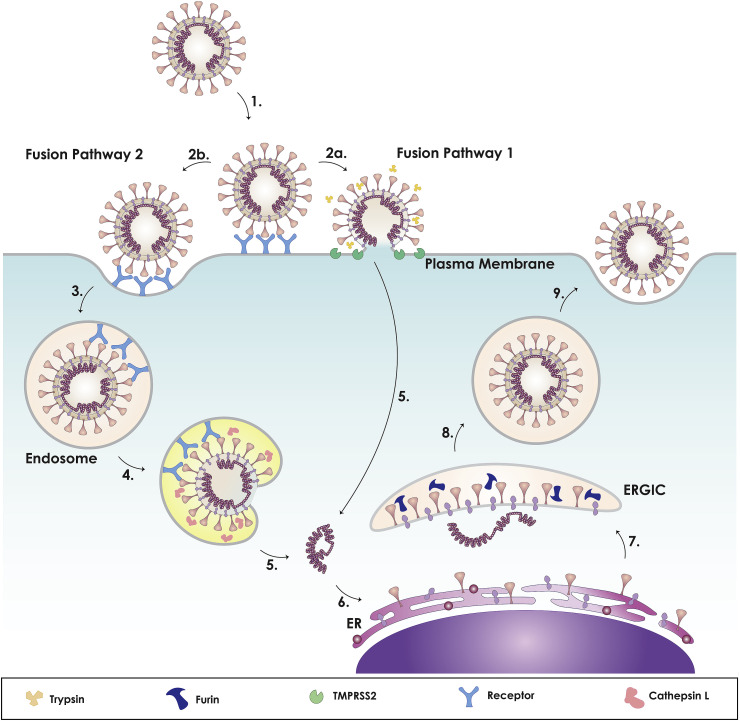
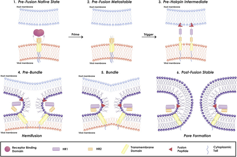
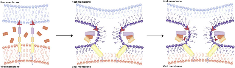
The coronavirus disease 2019 (COVID-19) pandemic has focused attention on the need to develop effective therapies against the causative agent, SARS-CoV-2, and also against other pathogenic coronaviruses (CoV) that have emerged in the past or might appear in future. Researchers are therefore focusing on steps in the CoV replication cycle that may be vulnerable to inhibition by broad-spectrum or specific antiviral agents. The conserved nature of the fusion domain and mechanism across the CoV family make it a valuable target to elucidate and develop pan-CoV therapeutics. In this article, we review the role of the CoV spike protein in mediating fusion of the viral and host cell membranes, summarizing the results of research on SARS-CoV, MERS-CoV, and recent peer-reviewed studies of SARS-CoV-2, and suggest that the fusion mechanism be investigated as a potential antiviral target. We also provide a supplemental file containing background information on the biology, epidemiology, and clinical features of all human-infecting coronaviruses, along with a phylogenetic tree of these coronaviruses.

Keywords: COVID-19; Fusion peptide; Middle east respiratory syndrome; SARS-CoV-2; Severe acute respiratory syndrome; Spike protein.


Figures

Fig. 1
Fig. 1
Coronavirus spike (S) protein. A. Cartoon figure of the CoV particle (top) and complete CoV viral genome (bottom). CoVs have a lipid envelope with three structural transmembrane proteins: spike (S), membrane (M), and envelope (E). The virus interior contains the viral genome encapsulated by the nucleocapsid (N) protein. The CoV single stranded genome encodes for 16 non-structural proteins, including the papain-like protease (PLpro), 3C-like protease (3CLpro), RNA-dependent RNA polymerase (RdRp), helicase (Hel), and exonuclease (ExoN). The subgenomic RNAs encode four structural proteins: spike (S; dark pink), envelope (E; dark blue), membrane (M; purple), and nucleocapsid (N; magenta) and a number of accessory proteins (Chan et al., 2020; de Wit et al., 2016). B. Cartoon figure of the CoV S protein trimer. C. The CoV S gene denoting the functional components of the protein. The CoV S protein is composed of the two subunits: S1 and S2, encompassing the major functional components: SP (signal peptide, pink); NTD (N-terminal domain; green), CTD (C-terminal domain; light blue), FP (fusion peptide; red), HR1 (heptad repeat 1; purple), HR2 (heptad repeat 2; orange), TM (transmembrane; yellow), and CP (cytoplasmic; dark blue). The S protein has two cleavage sites denoted with dark purple (S1/S2) and pink (S2’) arrows. D. Sequence alignment of S1/S2 cleavage site (dark purple arrow) and S2’ cleavage site (pink) between MERS-CoV, SARS-CoV, and SARS-CoV-2. E. Within the genome, the fusion peptide is highlighted, denoting the sequences from MERS-CoV FP and SARS-CoV FP. Red denotes the conserved residues between MERS-CoV, SARS-CoV, and SARS-CoV-2 FP sequences; blue denotes the SARS-CoV and SARS-CoV-2 FP conserved residues; green denotes the SARS-CoV and MERS-CoV FP conserved residues; purple denotes the MERS-CoV and SARS-CoV-2 conserved residues. The fusion peptide sequence of SARS-CoV-2 was determined by performing a pairwise alignment with MUSCLE through Geneious (version 2020.0.5). Amino acid sequence of the spike proteins was obtained from NCBI Genbank based on the following: SARS-CoV-2 (MN908947.3), MERS-CoV (AFS88936.1), SARS-CoV (AAP13441.1).
Fig. 2
Fig. 2
MERS-CoV (A), SARS-CoV (B), and SARS-CoV-2 (C) protein models. Models were built to show the predicted structure of the S1/S2, the S2′ cleavage site and the FP, which are not solved in cryo-EM structures. Trimers and monomers were modeled using SARS-CoV (PDB# 5X58) and MERS-CoV (PDB# 6Q05) structures using the methodology described in (Jaimes et al., 2020b). Color scheme is as described for Fig. 1.
Fig. 3
Fig. 3
Model of coronavirus dual entry pathway. This model depicts the two methods of viral entry: early pathway and late pathway. As the virus binds to its receptor (1), it can achieve entry via two routes: plasma membrane or endosome. For SARS-CoV: The presence of exogeneous and membrane bound proteases, such as trypsin and TMPRSS2, triggers the early fusion pathway (2a). Otherwise, it will be endocytosed (2b, 3). For MERS-CoV: If furin cleaved the S protein at S1/S2 during biosynthesis, exogeneous and membrane bound proteases, such as trypsin and TMPRSS2, will trigger early entry (2a). Otherwise, it will be cleaved at the S1/S2 site (2b) causing the virus to be endocytosed (3). For both: Within the endosome, the low pH activates cathepsin L (4), cleaving S2′ site, triggering the fusion pathway and releasing the CoV genome. Upon viral entry, copies of the genome are made in the cytoplasm (5), where components of the spike protein are synthesized in the rough endoplasmic reticulum (ER) (6). The structural proteins are assembled in the ER-Golgi intermediate compartment (ERGIC), where the spike protein can be pre-cleaved by furin, depending on cell type (7), followed by release of the virus from the cell (8, 9). For SARS-CoV-2: Studies currently show that SARS-CoV-2 can utilize membrane bound TMPRSS2 or endosomal cathepsin L for entry and that the S protein is processed during biosynthesis. Other factors that can influence the viral entry pathway are calcium and cholesterol (not shown).
Fig. 4
Fig. 4
Coronavirus viral fusion pathway model based on class I fusion protein understanding. The captions above the figure describe the state of the fusion protein, the captions below describe the state of the membranes. The S protein starts in the pre-fusion native state (1) and undergoes priming of the S1 subunit by relevant proteases to achieve the pre-fusion metastable state (2). Subsequent triggering by relevant proteases will enable the FP to insert in the host membrane and allow the S protein to form the pre-hairpin intermediate (3). The pre-hairpin begins to fold back on itself due to HR1 and HR2 interactions forming the pre-bundle (4), bundle (5), and eventual post-fusion stable (6) states. During the S protein foldback, the two membranes will approach each other until the outer leaflets merge (hemifusion) and eventually the inner leaflets merge (pore formation). Adapted from (White and Whittaker, 2016).
Fig. 5
Fig. 5
Model of major antiviral inhibitor pathway. This model depicts the inhibitory mechanism of a major CoV inhibitory peptide: HR2 peptide. Exogeneous HR2 peptides present during the CoV membrane fusion can competitively bind with CoV HR1. This prevents CoV HR2 from locking with HR1 and arrests the membrane fusion reaction, subsequently preventing pore formation.

References

    1. Alsaadi E.A.J., Neuman B.W., Jones I.M. A fusion peptide in the spike protein of MERS coronavirus. Viruses. 2019;11:825. doi: 10.3390/v11090825. - DOI - PMC - PubMed
    1. Andersen K.G., Rambaut A., Lipkin W.I., Holmes E.C., Garry R.F. The proximal origin of SARS-CoV-2. Nat. Med. 2020;89:44–48. doi: 10.1038/s41591-020-0820-9. - DOI - PMC - PubMed
    1. Barlan A., Zhao J., Sarkar M.K., Li K., McCray P.B., Perlman S., Gallagher T. Receptor variation and susceptibility to Middle East respiratory syndrome coronavirus infection. J. Virol. 2014;88:4953–4961. doi: 10.1128/JVI.00161-14. - DOI - PMC - PubMed
    1. Belouzard S., Chu V.C., Whittaker G.R. Activation of the SARS coronavirus spike protein via sequential proteolytic cleavage at two distinct sites. Proc. Natl. Acad. Sci. Unit. States Am. 2009;106:5871–5876. doi: 10.1073/PNAS.0809524106. - DOI - PMC - PubMed
    1. Belouzard S., Madu I., Whittaker G.R. Elastase-mediated activation of the severe acute respiratory syndrome coronavirus spike protein at discrete sites within the S2 domain * □ S downloaded from. J. Biol. Chem. 2010;285:22758–22763. doi: 10.1074/jbc.M110.103275. - DOI - PMC - PubMed

Publication types

Substances