

High-resolution Chest CT Features and Clinical Characteristics of Patients Infected with COVID-19 in Jiangsu, China
- PMID: 32272262
- PMCID: PMC7136866
- DOI: 10.1016/j.ijid.2020.04.003
High-resolution Chest CT Features and Clinical Characteristics of Patients Infected with COVID-19 in Jiangsu, China
Abstract
Background: A pneumonia associated with the coronavirus disease 2019 (COVID-19) recently emerged in China. It was recognized as a global health hazard.
Methods: 234 inpatients with COVID-19 were included. Detailed clinical data, chest HRCT basic performances and certain signs were recorded Ground-glass opacity (GGO), consolidation, fibrosis and air trapping were quantified. Both clinical types and CT stages were evaluated.
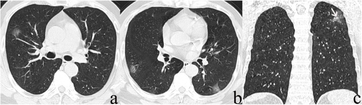
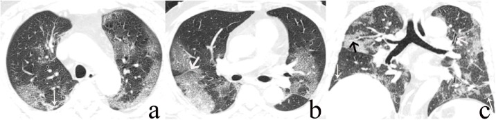
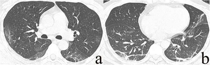
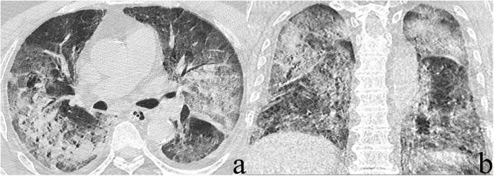
Results: Most patients (approximately 90%) were classified as common type and with epidemiologic history. Fever and cough were main symptoms. Chest CT showed abnormal attenuation in bilateral multiple lung lobes, distributed in the lower and/or periphery of the lungs (94.98%), with multiple shapes. GGO and vascular enhancement sign were most frequent seen, followed by interlobular septal thickening and air bronchus sign as well as consolidation, fibrosis and air trapping. There were significant differences in most of CT signs between different stage groups. The SpO2 and OI were decreased in stage IV, and the CT score of consolidation, fibrosis and air trapping was significantly lower in stage I (P<0.05). A weak relevance was between the fibrosis score and the value of PaO2 and SpO2 (P<0.05).
Conclusions: Clinical performances of patients with COVID-19, mostly with epidemiologic history and typical symptoms, were critical valuable in the diagnosis of the COVID-19. While chest HRCT provided the distribution, shape, attenuation and extent of lung lesions, as well as some typical CT signs of COVID-19 pneumonia.
Keywords: COVID-19; High-resolution CT (HRCT); SARS-CoV-2.
Copyright © 2020 The Author(s). Published by Elsevier Ltd.. All rights reserved.
Figures
Similar articles
-
Clinical characteristics and changes of chest CT features in 307 patients with common COVID-19 pneumonia infected SARS-CoV-2: A multicenter study in Jiangsu, China.Int J Infect Dis. 2020 Jul;96:157-162. doi: 10.1016/j.ijid.2020.05.006. Epub 2020 May 8. Int J Infect Dis. 2020. PMID: 32423888 Free PMC article.
-
[Clinical features and high resolution CT imaging evolution of coronavirus disease 2019].Zhonghua Jie He He Hu Xi Za Zhi. 2020 Jun 12;43(6):509-515. doi: 10.3760/cma.j.cn112147-20200214-00094. Zhonghua Jie He He Hu Xi Za Zhi. 2020. PMID: 32486557 Chinese.
-
Imaging and clinical features of patients with 2019 novel coronavirus SARS-CoV-2.Eur J Nucl Med Mol Imaging. 2020 May;47(5):1275-1280. doi: 10.1007/s00259-020-04735-9. Epub 2020 Feb 28. Eur J Nucl Med Mol Imaging. 2020. PMID: 32107577 Free PMC article.
-
Similarities and Differences of Early Pulmonary CT Features of Pneumonia Caused by SARS-CoV-2, SARS-CoV and MERS-CoV: Comparison Based on a Systemic Review.Chin Med Sci J. 2020 Sep 30;35(3):254-261. doi: 10.24920/003727. Chin Med Sci J. 2020. PMID: 32972503 Free PMC article.
-
Coronavirus Disease 2019 (COVID-19) CT Findings: A Systematic Review and Meta-analysis.J Am Coll Radiol. 2020 Jun;17(6):701-709. doi: 10.1016/j.jacr.2020.03.006. Epub 2020 Mar 25. J Am Coll Radiol. 2020. PMID: 32283052 Free PMC article.
Cited by
-
The importance of repeat testing in detecting coronavirus disease 2019 (COVID-19) in a coronary artery bypass grafting patient.J Card Surg. 2020 Jun;35(6):1342-1344. doi: 10.1111/jocs.14604. Epub 2020 May 12. J Card Surg. 2020. PMID: 32400044 Free PMC article. Review.
-
Medical imaging and computational image analysis in COVID-19 diagnosis: A review.Comput Biol Med. 2021 Aug;135:104605. doi: 10.1016/j.compbiomed.2021.104605. Epub 2021 Jun 23. Comput Biol Med. 2021. PMID: 34175533 Free PMC article. Review.
-
Diagnostic features of SARS-COVID-2-positive patients: A rapid review and meta-analysis.J Clin Nurs. 2021 Jul;30(13-14):1826-1837. doi: 10.1111/jocn.15688. Epub 2021 Feb 17. J Clin Nurs. 2021. PMID: 33527510 Free PMC article. Review.
-
Thoracic imaging tests for the diagnosis of COVID-19.Cochrane Database Syst Rev. 2022 May 16;5(5):CD013639. doi: 10.1002/14651858.CD013639.pub5. Cochrane Database Syst Rev. 2022. PMID: 35575286 Free PMC article.
-
A Case of Spontaneous Pneumothorax 21 Days After Diagnosis of Coronavirus Disease 2019 (COVID-19) Pneumonia.Am J Case Rep. 2020 Aug 15;21:e925787. doi: 10.12659/AJCR.925787. Am J Case Rep. 2020. PMID: 32798215 Free PMC article.
References
-
- Ksiazek T.G., Erdman D., Goldsmith C.S., Zaki S.R., Peret T., Emery S., Tong S., Urbani C., Comer J.A., Lim W., Rollin P.E., Dowell S.F., Ling A.E., Humphrey C.D., Shieh W.J., Guarner J., Paddock C.D., Rota P., Fields B., DeRisi J., Yang J.Y., Cox N., Hughes J.M., LeDuc J.W., Bellini W.J., Anderson L.J., SARS Working Group A novel coronavirus associated with severe acute respiratory syndrome. N Engl J Med. 2003;348:1953–1966. - PubMed
-
- K Kuiken T., Fouchier R.A., Schutten M., Rimmelzwaan G.F., van Amerongen G., van Riel D., Laman J.D., de Jong T., van Doornum G., Lim W., Ling A.E., Chan P.K., Tam J.S., Zambon M.C., Gopal R., Drosten C., van der Werf S., Escriou N., Manuguerra J.C., Stöhr K., Peiris J.S., Osterhaus A.D. Lancet. 2003;362:263–270. - PMC - PubMed
-
- de Groot R.J., Baker S.C., Baric R.S., Brown C.S., Drosten C., Enjuanes L., Fouchier R.A., Galiano M., Gorbalenya A.E., Memish Z.A., Perlman S., Poon L.L., Snijder E.J., Stephens G.M., Woo P.C., Zaki A.M., Zambon M., Ziebuhr J. Middle East respiratory syndrome coronavirus (MERS-CoV): announcement of the Coronavirus Study Group. J Virol. 2013;87:7790–7792. - PMC - PubMed
-
- Huang C., Wang Y., Li X., Ren L., Zhao J., Hu Y., Zhang L., Fan G., Xu J., Gu X., Cheng Z., Yu T., Xia J., Wei Y., Wu W., Xie X., Yin W., Li H., Liu M., Xiao Y., Gao H., Guo L., Xie J., Wang G., Jiang R., Gao Z., Jin Q., Wang J., Cao B. Clinical features of patients infected with 2019 novel coronavirus in Wuhan. China.Lancet. 2020;395:497–506. - PMC - PubMed
-
- Chung M., Bernheim A., Mei X., Zhang N., Huang M., Zeng X., Cui J., Xu W., Yang Y., Fayad Z.A., Jacobi A., Li K., Li S., Shan H. CT Imaging Features of 2019 Novel Coronavirus (COVID-19) Radiology. 2020:200230. [Preprint]. Available from: https://doi.org/10.1148/radiol.2020200230. - PMC - PubMed
MeSH terms
LinkOut - more resources
Full Text Sources
Other Literature Sources
Medical
Miscellaneous
