. 2020 Jun;215(2):373-378.
doi: 10.1534/genetics.120.303182. Epub 2020 Apr 9.

Oocyte Elimination Through DNA Damage Signaling from CHK1/CHK2 to p53 and p63

Affiliations

Oocyte Elimination Through DNA Damage Signaling from CHK1/CHK2 to p53 and p63

Vera D Rinaldi et al. Genetics. 2020 Jun.

Abstract

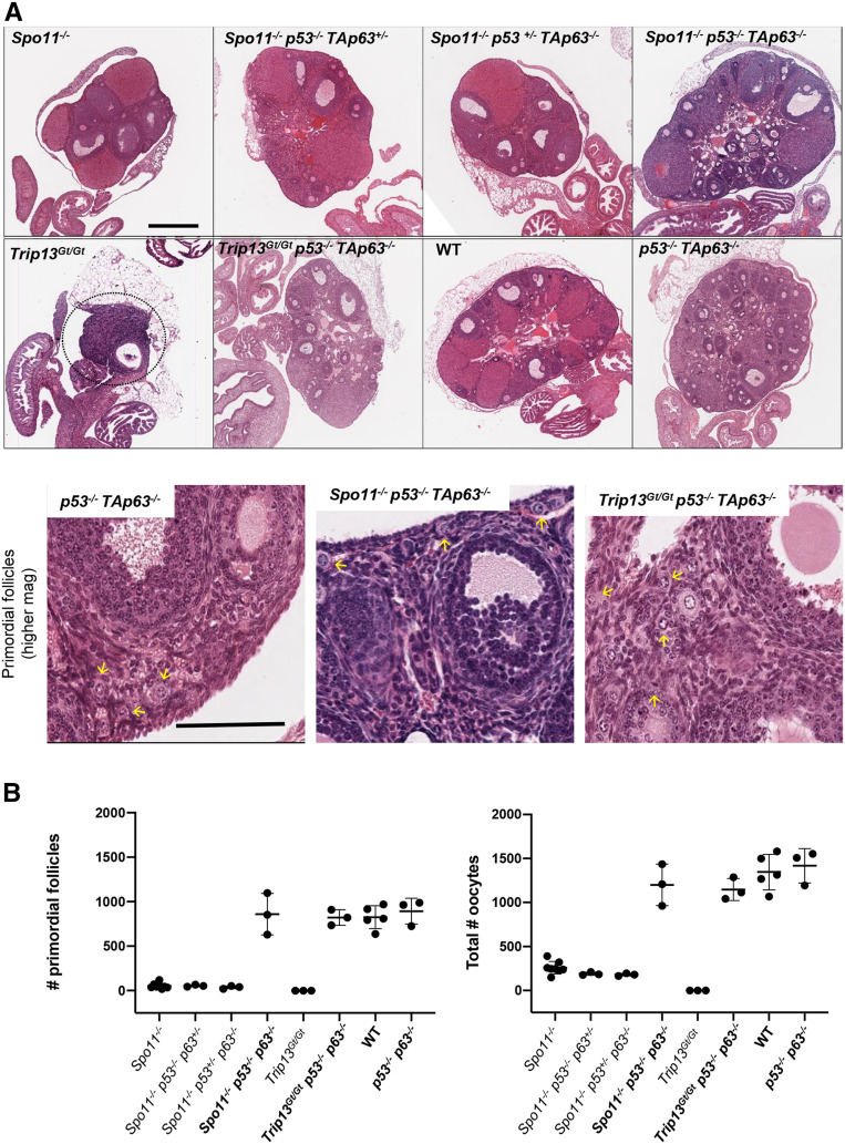
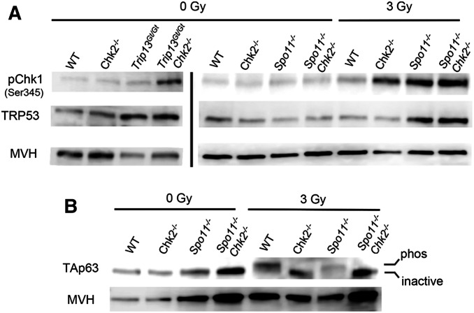
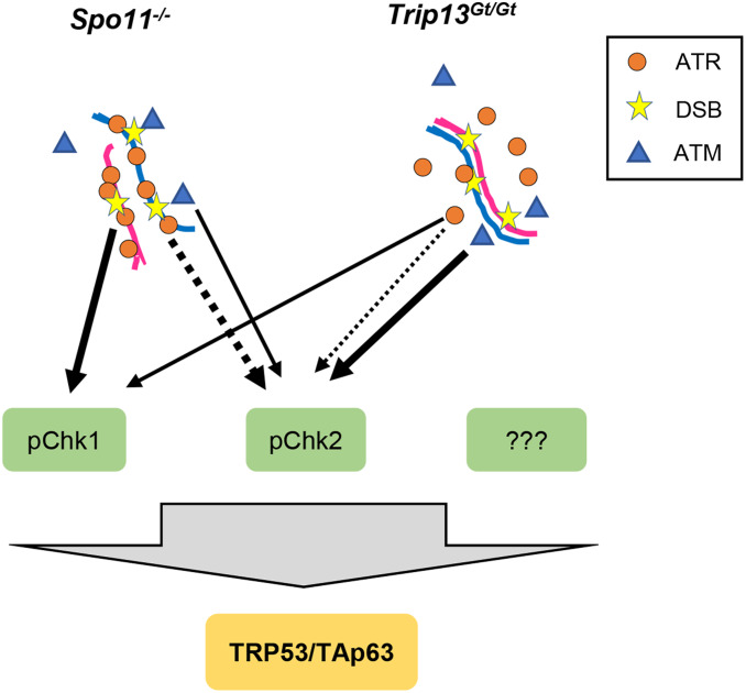
Eukaryotic organisms have evolved mechanisms to prevent the accumulation of cells bearing genetic aberrations. This is especially crucial for the germline, because fecundity and fitness of progeny would be adversely affected by an excessively high mutational incidence. The process of meiosis poses unique problems for mutation avoidance because of the requirement for SPO11-induced programmed double-strand breaks (DSBs) in recombination-driven pairing and segregation of homologous chromosomes. Mouse meiocytes bearing unrepaired meiotic DSBs or unsynapsed chromosomes are eliminated before completing meiotic prophase I. In previous work, we showed that checkpoint kinase 2 (CHK2; CHEK2), a canonical DNA damage response protein, is crucial for eliminating not only oocytes defective in meiotic DSB repair (e.g., Trip13Gt mutants), but also Spo11-/- oocytes that are defective in homologous chromosome synapsis and accumulate a threshold level of spontaneous DSBs. However, rescue of such oocytes by Chk2 deficiency was incomplete, raising the possibility that a parallel checkpoint pathway(s) exists. Here, we show that mouse oocytes lacking both p53 (TRP53) and the oocyte-exclusive isoform of p63, TAp63, protects nearly all Spo11-/- and Trip13Gt/Gt oocytes from elimination. We present evidence that checkpoint kinase I (CHK1; CHEK1), which is known to signal to TRP53, also becomes activated by persistent DSBs in oocytes, and to an increased degree when CHK2 is absent. The combined data indicate that nearly all oocytes reaching a threshold level of unrepaired DSBs are eliminated by a semiredundant pathway of CHK1/CHK2 signaling to TRP53/TAp63.

Keywords: checkpoints; meiosis; mouse; oocytes; transducer kinases.


Figures

Figure 1
Figure 1
Rescue of SPO11- and TRIP13-deficient oocytes by compound deletion of p53 and TAp63. (A) Hematoxylin and eosin stained ovaries from 2-month-old mice of the indicated genotypes. The top two rows are images are from histological sections through the approximate center of the ovaries. Bar, 500 µm. The dashed circle indicates the residual Trip13Gt/Gt ovary. The bottom row shows higher magnification images of selected genotypes. Bar, 100 µm. Yellow arrows indicate examples of primordial follicles. (B) Oocyte quantification. To the left is the quantification of primordial follicles [P < 0.0001 for all oocyte rescued (in bold) genotypes compared to nonrescued genotypes; P = 0.92 for oocyte rescued genotypes vs. WT and Trp53−/− TAp63−/− ovaries]. To the right are total oocytes (from all stages of follicles) from individual ovaries (P < 0.0001 for all oocyte rescued genotypes compared to nonrescued genotypes; P = 0.33 for oocyte rescued genotypes vs. WT and Trp53−/− TAp63−/− ovaries). Oocyte quantification data are presented in Table S1. Genotype abbreviations are as follows: TAp63 is abbreviated as p63; WT, wild type.
Figure 2
Figure 2
Increased CHK1 activation and p53 stabilization, but not TAp63 activation, in CHK2-deficient oocytes. (A) CHK1 phosphorylation in oocytes is stimulated by induced or meiotic DSBs. Shown are Western blots probed with indicated antibodies. Each lane contains total protein extracted from four ovaries (postnatal day 3–5) that were either exposed or not to 3 Gy of ionizing radiation (IR). Ovaries were harvested for protein extraction 3 hr post-IR. The blots on the left, separated by a vertical bar from those on the right, were from a different blot and different protein samples and mice. The same two blots (left and right) were stripped and reprobed sequentially with the three antibodies. A biological replicate is shown in Figure S1. Note that the decreased MVH levels in Trip13Gt/Gt ovaries is due to reduction in oocytes. (B) Activation of the TAp63 isoform is dependent on DNA damage and CHK2 signaling, not asynapsis. Shown is a Western blot probed sequentially for TAp63 and the germ cell marker MVH. Each lane contains protein extracted from ovaries as described in A. An upward shift in the band indicates the presence of the active (phosphorylated) vs. inactive TAp63. A biological replicate is shown in Figure S1.
Figure 3
Figure 3
Model of checkpoint signaling in mouse oocytes. We propose that all DSB damage signaling in oocytes requires activation of TRP53 and TAp63 for complete oocyte elimination. The dashed lines represent noncanonical phosphorylation of CHK2 by ATR, and the thickness of all lines represents the relative amounts of activation in the two indicated mutant situations. We propose that in highly asynaptic Spo11 mutant oocytes, the “preloading” of ATR as part of the MSUC response leads it to play a larger role in signaling to CHK1 and CHK2 than under situations in which DSBs occur on synapsed chromosomes.

References

    1. Abe H., Alavattam K. G., Kato Y., Castrillon D. H., Pang Q. et al. , 2018. CHEK1 coordinates DNA damage signaling and meiotic progression in the male germline of mice. Hum. Mol. Genet. 27: 1136–1149. 10.1093/hmg/ddy022 - DOI - PMC - PubMed
    1. Balmus G., Zhu M., Mukherjee S., Lyndaker A. M., Hume K. R. et al. , 2012. Disease severity in a mouse model of ataxia telangiectasia is modulated by the DNA damage checkpoint gene Hus1. Hum. Mol. Genet. 21: 3408–3420. 10.1093/hmg/dds173 - DOI - PMC - PubMed
    1. Baudat F., Manova K., Yuen J. P., Jasin M., and Keeney S., 2000. Chromosome synapsis defects and sexually dimorphic meiotic progression in mice lacking. Mol. Cell 6: 989–998. 10.1016/S1097-2765(00)00098-8 - DOI - PubMed
    1. Bolcun-Filas E., Rinaldi V. D., White M. E., and Schimenti J. C., 2014. Reversal of female infertility by Chk2 ablation reveals the oocyte DNA damage checkpoint pathway. Science 343: 533–536. 10.1126/science.1247671 - DOI - PMC - PubMed
    1. Carofiglio F., Inagaki A., de Vries S., Wassenaar E., Schoenmakers S. et al. , 2013. SPO11-independent DNA repair foci and their role in meiotic silencing. PLoS Genet. 9: e1003538 10.1371/journal.pgen.1003538 - DOI - PMC - PubMed

Publication types

MeSH terms