

Suspected Metallic Embolization Distal to Coiled Intracranial Aneurysms Detectable by Susceptibility-Weighted MR Imaging
- PMID: 32273325
- PMCID: PMC7144647
- DOI: 10.3174/ajnr.A6506
Suspected Metallic Embolization Distal to Coiled Intracranial Aneurysms Detectable by Susceptibility-Weighted MR Imaging
Abstract
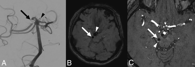
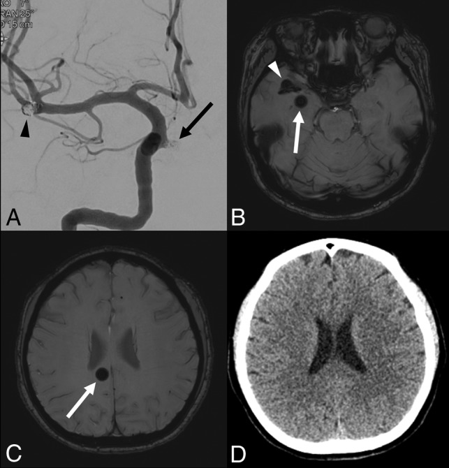
Background and purpose: After endovascular coiling of intracranial aneurysms, round dark parenchymal lesions believed to be particulate metal are sometimes encountered in MR imaging studies of the brain. We used SWI to assess the frequency of such occurrences, in addition to exploring likely causes and clinical implications.
Materials and methods: We reviewed 700 MR imaging studies performed between September 2018 and March 2019 at our institution as follow-up monitoring of coiled intracranial aneurysms. Any sizeable (>5 mm) rounded dark-signal lesions encountered were presumed to be metallic. The magnitudes and locations of such lesions were recorded. In patients with these lesions, pertinent procedural documentation was screened for devices used, including coils, microcatheters, microguidewires, and stents. Medical records were also examined to determine whether any related symptoms ensued.
Results: Twenty patients (2.8%) exhibited a total of 25 lesions on SWI. Diameters ranged from 5 to 11 mm (median, 8 mm). All except 2 lesions were located in brain regions downstream from aneurysms, but all lesions occupied vascular territories of vessels used to place guiding catheters. Other than the Synchro 14, which was routinely deployed, no device was regularly used in patients with SWI-detectable lesions; and none of the affected patients developed focal neurologic symptoms as a consequence.
Conclusions: Although the origins remain unclear, distal embolization of particulate metal distal to coiled cerebral aneurysms is occasionally observed on follow-up MR imaging studies. Such lesions, however, seem to have no apparent clinical impact.
© 2020 by American Journal of Neuroradiology.
Figures
Similar articles
-
Suspected Metallic Embolism following Endovascular Treatment of Intracranial Aneurysms.AJNR Am J Neuroradiol. 2016 Sep;37(9):1696-9. doi: 10.3174/ajnr.A4804. Epub 2016 Apr 21. AJNR Am J Neuroradiol. 2016. PMID: 27102315 Free PMC article.
-
Evaluation of ischemic lesion prevalence after endovascular treatment of intracranial aneurysms, as documented by 3-T diffusion-weighted imaging: a 2-year, single-center cohort study.J Neurosurg. 2018 Apr;128(4):982-991. doi: 10.3171/2016.11.JNS161020. Epub 2017 Jun 9. J Neurosurg. 2018. PMID: 28598274
-
Delayed enhancing lesions after coil embolization of aneurysms: clinical experience and benchtop analyses.J Neurointerv Surg. 2017 Dec;9(12):1243-1247. doi: 10.1136/neurintsurg-2016-012833. Epub 2017 Jan 6. J Neurointerv Surg. 2017. PMID: 28062804
-
Kissing-Y stenting for endovascular treatment of complex wide necked bifurcation aneurysms using Acandis Acclino stents: results and literature review.J Neurointerv Surg. 2016 Apr;8(4):386-95. doi: 10.1136/neurintsurg-2015-011691. Epub 2015 May 18. J Neurointerv Surg. 2016. PMID: 25987589 Review.
-
Endovascular methods for the treatment of intracranial cerebral aneurysms.Neuroimaging Clin N Am. 2013 Nov;23(4):563-91. doi: 10.1016/j.nic.2013.03.007. Epub 2013 May 25. Neuroimaging Clin N Am. 2013. PMID: 24156851 Review.
Cited by
-
Fragmentation of Hydrophilic Guidewire Coatings During Neuroendovascular Therapy.Clin Neuroradiol. 2023 Sep;33(3):793-799. doi: 10.1007/s00062-023-01283-1. Epub 2023 Apr 25. Clin Neuroradiol. 2023. PMID: 37185670
-
Clinically Silent Microinfarct Incidence and Risk Factors After Treatment of Unruptured Intracranial Aneurysms with Hydrophilic Polymer-Coated Flow Diverters.Clin Neuroradiol. 2025 Feb 14. doi: 10.1007/s00062-025-01497-5. Online ahead of print. Clin Neuroradiol. 2025. PMID: 39953138
-
Hydrophilic polymer coating delamination during neurointerventional treatment after microcatheter withdrawal: particulate identification through attenuated total reflection Fourier-transform infrared spectroscopy.Front Neurol. 2025 Jan 15;15:1479375. doi: 10.3389/fneur.2024.1479375. eCollection 2024. Front Neurol. 2025. PMID: 39882360 Free PMC article.
References
-
- Molyneux A, Kerr R, Stratton I, et al. . International Subarachnoid Aneurysm Trial (ISAT) of neurosurgical clipping versus endovascular coiling in 2143 patients with ruptured intracranial aneurysms: a randomised trial. Lancet 2002;360:1267–74 10.1016/S0140-6736(02)11314-6 10.1016/s0140-6736(02)11314-6 - DOI - DOI - PubMed
MeSH terms
Substances
LinkOut - more resources
Full Text Sources
Medical