. 2020 Apr 28;117(17):9537-9545.
doi: 10.1073/pnas.1916054117. Epub 2020 Apr 9.

PSGL-1 restricts HIV-1 infectivity by blocking virus particle attachment to target cells

Affiliations

PSGL-1 restricts HIV-1 infectivity by blocking virus particle attachment to target cells

Yajing Fu et al. Proc Natl Acad Sci U S A. .

Abstract

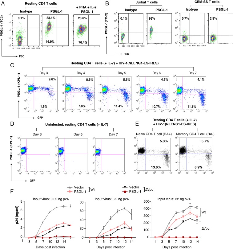
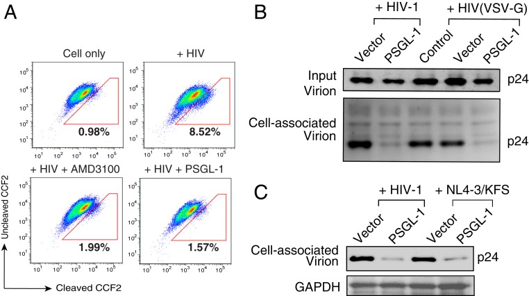
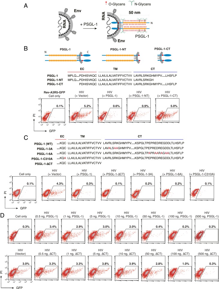
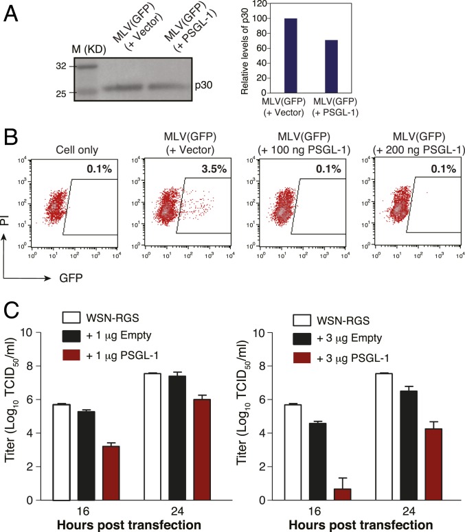
P-selectin glycoprotein ligand-1 (PSGL-1) is a dimeric, mucin-like, 120-kDa glycoprotein that binds to P-, E-, and L-selectins. PSGL-1 is expressed primarily on the surface of lymphoid and myeloid cells and is up-regulated during inflammation to mediate leukocyte tethering and rolling on the surface of endothelium for migration into inflamed tissues. Although it has been reported that PSGL-1 expression inhibits HIV-1 replication, the mechanism of PSGL-1-mediated anti-HIV activity remains to be elucidated. Here we report that PSGL-1 in virions blocks the infectivity of HIV-1 particles by preventing the binding of particles to target cells. This inhibitory activity is independent of the viral glycoprotein present on the virus particle; the binding of particles bearing the HIV-1 envelope glycoprotein or vesicular stomatitis virus G glycoprotein or even lacking a viral glycoprotein is impaired by PSGL-1. Mapping studies show that the extracellular N-terminal domain of PSGL-1 is necessary for its anti-HIV-1 activity, and that the PSGL-1 cytoplasmic tail contributes to inhibition. In addition, we demonstrate that the PSGL-1-related monomeric E-selectin-binding glycoprotein CD43 also effectively blocks HIV-1 infectivity. HIV-1 infection, or expression of either Vpu or Nef, down-regulates PSGL-1 from the cell surface; expression of Vpu appears to be primarily responsible for enabling the virus to partially escape PSGL-1-mediated restriction. Finally, we show that PSGL-1 inhibits the infectivity of other viruses, such as murine leukemia virus and influenza A virus. These findings demonstrate that PSGL-1 is a broad-spectrum antiviral host factor with a unique mechanism of action.

Keywords: CD43; HIV-1; Nef; PSGL-1; Vpu.


Conflict of interest statement

Competing interest statement: Two provisional patent applications pertaining to the results presented in this paper have been filed by George Mason University.

Figures

Fig. 1.
Fig. 1.
PSGL-1 is expressed on human CD4 T cells and exerts an anti-HIV activity. (A) Peripheral blood resting CD4 T cells were purified by negative selection, activated with PHA+IL-2, or left unstimulated. Cell surface PSGL-1 expression was analyzed by flow cytometry. (B) Jurkat and CEM-SS cells were similarly stained for surface PSGL-1. (CE) Down-regulation of PSGL-1 by HIV-1 in primary CD4 T cells. Primary resting CD4 T cells were infected with NLENG1-ES-IRES, a GFP reporter virus. Following infection, cells were washed and cultured in complete medium plus IL-7 (2 ng/mL) to permit low-level viral replication. Surface PSGL-1 expression was analyzed at the indicated days. (C) Percentages of the GFP+ or GFP cells with low or high PSGL-1 staining in each panel. PSGL-1 down-regulation was observed only in the HIV-infected (GFP+) cell population. (D) For controls, uninfected cells were similarly cultured in IL-7, and surface PSGL-1 expression was analyzed at the indicated days. Culturing of resting CD4 T cells in IL-7 did not lead to PSGL-1 down-regulation. (E) Cells were also stained for surface PSGL-1 expression on CD45RA+ (naïve) and CD45RA (memory) CD4 T cells on day 7 and analyzed by flow cytometry. (F) HeLa JC.53 cells were stably transfected with PSGL-1 or empty vector DNA and drug-selected to obtain stably transfected cells. These cells were then infected with three different inputs of HIV-1(NL4-3)WT or HIV-1(∆Vpu). Viral replication was quantified by p24 release.
Fig. 2.
Fig. 2.
PSGL-1 does not block viral release but inactivates virion infectivity. (A) HeLa JC.53 cells were stably transfected with PSGL-1 or empty vector DNA and drug-selected to obtain stably PSGL-1–expressing cells. Cells were then infected with the single-round vector HIV-1 (gp160) for single-round infection. Viral replication was quantified by p24 release. The black and red bars represent empty vector and PSGL-1 vector-transfected cells, respectively. (BE) HEK293T cells were cotransfected with HIV(NL4-3) DNA (1 μg) plus different amounts of PSGL-1 expression vector. Viral p24 release was quantified at 48 h (B). Cells were also lysed and analyzed by Western blot for intracellular PSGL-1 and HIV-1 proteins (C). Extracellular virion p24 was also analyzed by Western blot (D), and the relative ratio of extracellular and intracellular p24 was plotted (E). The black and red bars represent empty vector and PSGL-1 vector cotransfected cells, respectively. (F and G) Virions released from HEK293T cells cotransfected with HIV(NL4-3) DNA (1 μg) plus PSGL-1 DNA (0.5 to 400 ng) were harvested at 48 h and normalized for p24, and viral infectivity was quantified by infecting the T cell line-derived Rev-A3R5-GFP indicator cell line. HIV-1 replication was quantified by GFP expression. Shown are the percentages of GFP+ cells at 48 h postinfection. (G) The PSGL-1 dose-dependent inhibition curve was plotted using results from three independent experiments. (H) PSGL-1 blocks the infectivity of VSV-G-pseudotyped HIV-1. Virus particles produced in the presence of PSGL-1 or the empty vector were assayed for infectivity using TZM-bl cells. (IK) shRNA knockdown of PSGL-1 enhances virion infectivity. Jurkat cells were transduced with a lentiviral vector expressing shRNA against PSGL-1 (shRNA PSGL-1) or a nontarget sequence (shRNA NTC). PSGL-1 surface expression was quantified (I) at 12 d posttransduction and puromycin selection. Cells were also electroporated with HIV-1(NL4-3) DNA, and viral replication in knockdown Jurkat cells was quantified by measuring p24 in the supernatant (J). To quantify HIV infectivity, virions were harvested at 48 h postelectroporation and used to infect Rev-A3R5-GFP cells, using an equal amount of p24 for infection (K).
Fig. 3.
Fig. 3.
PSGL-1 blocks virion attachment to target cells. (A) Virions produced from HEK293T cells cotransfected with PSGL-1 expression vector plus HIV-1(NL4-3) were used for an entry assay. HIV-1 virions similarly produced in the presence of an empty vector served as controls. Equal p24 was used for the assay. The entry inhibitor AMD3100 was also used as a control to block virus entry. The percentages of cells with cleaved CCF2 are shown. (B) Virions produced in the presence of PSGL-1 or the empty vector were assayed for attachment to target HeLa JC.53 cells at 4 °C for 2 h. Cells were washed and then analyzed by Western blot for bound p24. (C) PSGL-1 blocks Env(−) HIV-1 particle attachment to target cells. WT HIV-1 or Env(−) HIV-1 [(NL4-3)/KFS] virions were produced in the presence of PSGL-1 or the empty vector, and viral particles were assayed for attachment to target HeLa JC.53 cells.
Fig. 4.
Fig. 4.
The extracellular N-terminal domain of PSGL-1 is required to block HIV-1 infectivity. (A) Hypothetical model by which virion incorporation of PSGL-1 restricts virion infectivity. Based on this model, incorporation of the heavily glycosylated and elongated PSGL-1 on viral particles may interfere with virion binding to target cells. (B) PSGL-1 domains involved in blocking HIV-1 infectivity. HEK293T cells were cotransfected with HIV(NL4-3) DNA (1 μg) plus vectors expressing PSGL-1 or PSGL-1 truncation mutants PSGL-1-NT or PSGL-1-CT (500 ng). Virions were harvested at 48 h posttransfection and normalized for p24, and viral infectivity was quantified by infecting Rev-A3R5-GFP indicator cells. HIV-1 replication was quantified by GFP expression. (C) PSGL-1 intracellular domain mutants PSGL-1-∆CT, PSGL-1-3A, and PSGL-1-6A and the dimerization mutant PSGL-1-C310A were similarly tested. EC, extracellular domain; TM, transmembrane domain; CT, cytoplasmic tail. (D) The PSGL-1-∆CT mutant displays reduced antiviral activity relative to WT PSGL-1. HEK293T cells were cotransfected with HIV(NL4-3) DNA (1 μg) plus various amounts of PSGL-1 or PSGL-1-∆CT (0.5 to 500 ng). Virions were harvested at 48 h and normalized for p24, and their infectivity was measured in Rev-A3R5-GFP indicator cells.
Fig. 5.
Fig. 5.
PSGL-1 restricts MLV and influenza A virus infectivity. (A) HEK293T cells were cotransfected with an MLV helper vector, pSV-ψ-MLV-env, pRetroQ-AcGFP-N1, pHCMV-G, plus a PSGL-1 expression vector or an empty control vector. Virions were harvested and quantified by Western blot analysis using an anti-MLV p30 (CA) protein antibody. (B) MLV virions were harvested, and viral infectivity was quantified by infecting HEK293T cells and measuring GFP expression. (C) Eight vectors expressing each of the segments of the influenza A/WSN/33 (H1N1) genome were cotransfected with a PSGL-1 expression vector into HEK293T-MDCK cells. Viral particles were harvested at 16 and 24 h after cotransfection, and virion infectivity was quantified by a TCID50 assay.

References

    1. Sako D., et al. , Expression cloning of a functional glycoprotein ligand for P-selectin. Cell 75, 1179–1186 (1993). - PubMed
    1. Guyer D. A., et al. , P-selectin glycoprotein ligand-1 (PSGL-1) is a ligand for L-selectin in neutrophil aggregation. Blood 88, 2415–2421 (1996). - PubMed
    1. Goetz D. J., et al. , Isolated P-selectin glycoprotein ligand-1 dynamic adhesion to P- and E-selectin. J. Cell Biol. 137, 509–519 (1997). - PMC - PubMed
    1. Laszik Z., et al. , P-selectin glycoprotein ligand-1 is broadly expressed in cells of myeloid, lymphoid, and dendritic lineage and in some nonhematopoietic cells. Blood 88, 3010–3021 (1996). - PubMed
    1. Fujimoto T. T., et al. , Expression and functional characterization of the P-selectin glycoprotein ligand-1 in various cells. Int. J. Hematol. 64, 231–239 (1996). - PubMed

Publication types

Substances

LinkOut - more resources