. 2020 Apr 10;368(6487):186-189.
doi: 10.1126/science.aau6481.

Dendritic cell-derived hepcidin sequesters iron from the microbiota to promote mucosal healing

Affiliations

Dendritic cell-derived hepcidin sequesters iron from the microbiota to promote mucosal healing

Nicholas J Bessman et al. Science. .

Abstract

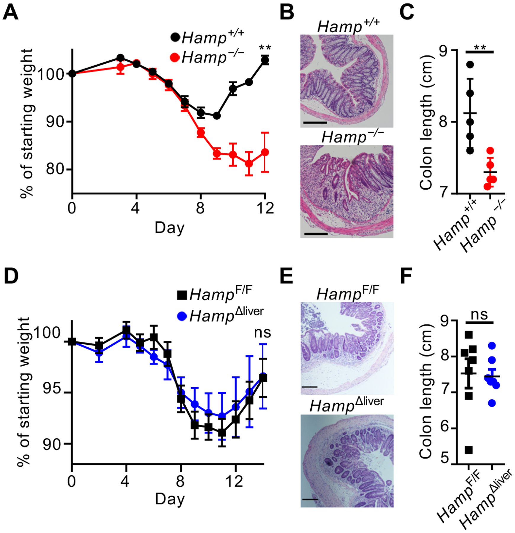
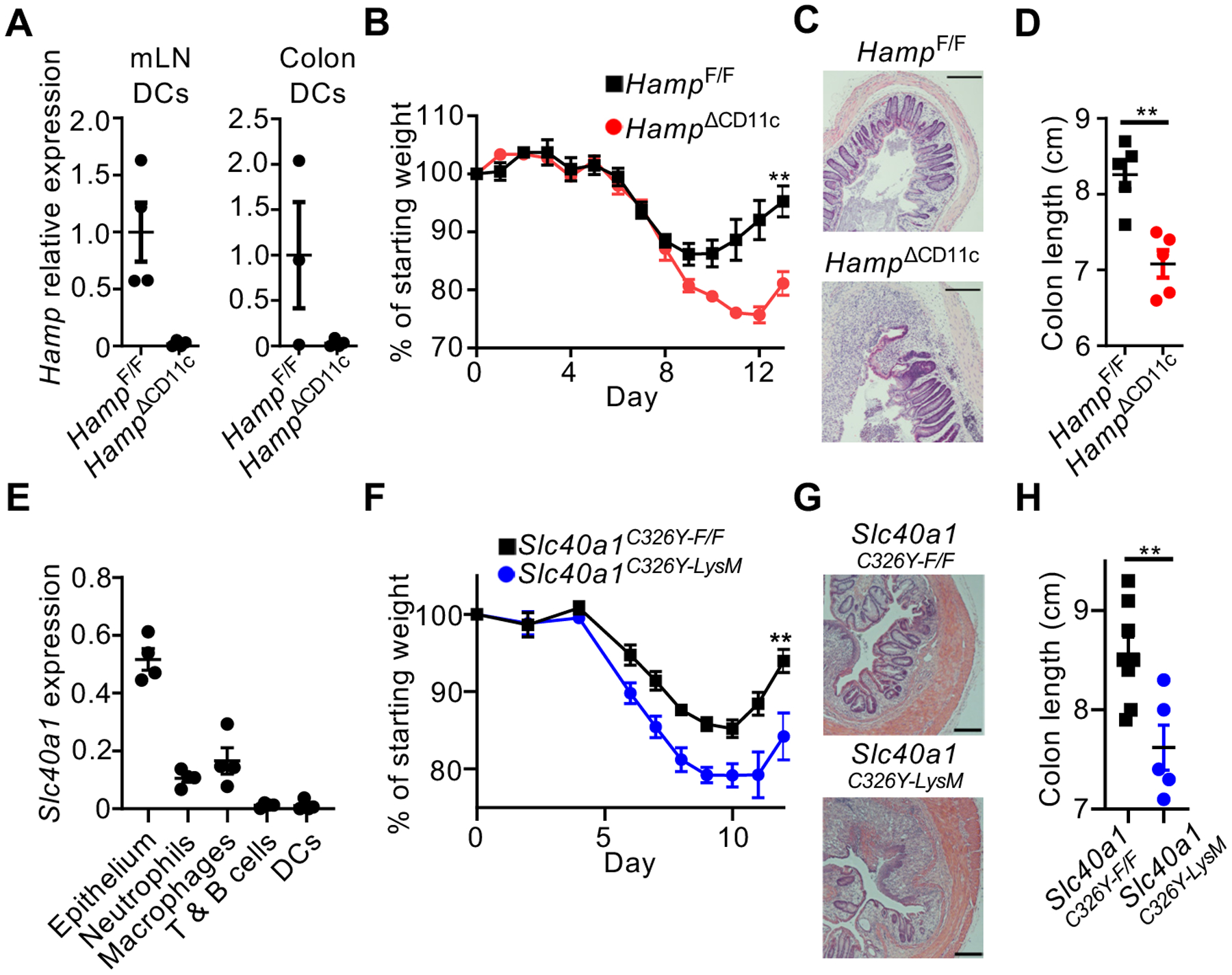
Bleeding and altered iron distribution occur in multiple gastrointestinal diseases, but the importance and regulation of these changes remain unclear. We found that hepcidin, the master regulator of systemic iron homeostasis, is required for tissue repair in the mouse intestine after experimental damage. This effect was independent of hepatocyte-derived hepcidin or systemic iron levels. Rather, we identified conventional dendritic cells (cDCs) as a source of hepcidin that is induced by microbial stimulation in mice, prominent in the inflamed intestine of humans, and essential for tissue repair. cDC-derived hepcidin acted on ferroportin-expressing phagocytes to promote local iron sequestration, which regulated the microbiota and consequently facilitated intestinal repair. Collectively, these results identify a pathway whereby cDC-derived hepcidin promotes mucosal healing in the intestine through means of nutritional immunity.


Conflict of interest statement

Competing interests: G.F.S holds stock and is a member of an advisory board for Celsius Therapeutics Inc. T.A. is an employee of Amgen, Inc.. The other authors declare no competing interests.

Figures

Fig. 1.
Fig. 1.. Extra-hepatic hepcidin promotes mucosal healing.
Mice were given DSS for 7 days, and disease and recovery was monitored by weight loss (A), H&E staining of distal colon (B), and colon shortening (C) at day 12. Mice were given DSS for 9 days and recovery was monitored by weight loss (D), H&E staining of the distal colon (E), and colon shortening (F). All scale bars are 200 μm. Data in (A-C, E) are representative of n=3–5 mice per group replicated in two or more independent experiments, and data in D and F are pooled from two independent experiments with n=3–4. Data are shown as the mean ± SEM. Statistics comparing groups used unpaired two-tailed Student’s t-test (**:p<0.01; ***:p<0.001; ****:p<0.0001). In (A) and (D), weights at sacrifice, normalized to starting weight, were analyzed by unpaired two-tailed Student’s t-test.
Fig. 2.
Fig. 2.. Conventional dendritic cells are a source of hepcidin in the inflamed intestine.
Hepcidin expression was analyzed by qPCR in naïve mouse tissue (A). Mice were provided DSS for 7 days, colon lamina propria myeloid cells were sorted as noted (B), and hepcidin expression was analyzed by qPCR (C). cDC2s sorted from spleen were stimulated and hepcidin expression was analyzed (D). Hepcidin expression was quantified from intestinal biopsies of humans (E). Lamina propria cells from the inflamed ileum of pediatric CD patients were analyzed for hepcidin protein (F-G). For (A, C, and D), representative data with n=3–5 per group are shown, and data were replicated in at least two independent trials. In (E), n=5 for the healthy group and n=21 for the UC and CD groups. In (F), representative histograms are shown. In (G), four independent patients were tested and all data was pooled. All data are shown as mean ± SEM. In (A, D, and E) data were analyzed by unpaired two-tailed Student’s t-test; in (C), data were analyzed by the Mann–Whitney U test; in (G), data were analyzed by one-way ANOVA with Tukey’s multiple comparisons test (*:p<0.05; **:p<0.01; ***:p<0.001).
Fig. 3.
Fig. 3.. Dendritic cell-derived hepcidin acts on ferroportin-expressing phagocytes to facilitate mucosal healing.
Hepcidin expression was determined by qPCR in mice exposed to DSS for 7 days (A). Mice were given DSS for 8 days, and recovery was monitored by weight change (B), H&E staining of distal colon (C), and colon shortening (D). Sort-purified cells from the naïve mouse colon were analyzed for Slc40a1 expression by qPCR (E). Mice were given DSS for 7 days and recovery was monitored by weight change (F), H&E staining of distal colon (G), and colon shortening (H). All scale bars are 200 μm. All data are shown as mean ± SEM. Data in (D) and (H) were analyzed by unpaired two-tailed Student’s t-test. In (B) and (F), weights at sacrifice, normalized to starting weight, were analyzed by unpaired two-tailed Student’s t-test. For all statistical tests, *:p<0.05; **:p<0.01; ***:p<0.001; ****:p<0.0001. Data in (A-D) are representative of at least two independent experiments with n=3–5 per group. Data in (F) and (H) are pooled from, and data in (J) is representative of, three independent experiments with n=1–3 per group.
Fig. 4.
Fig. 4.. Dendritic cell-derived hepcidin sequesters iron to shape the intestinal microbiota.
Mice were exposed DSS for 7 days. Whole ceca tissues were analyzed for iron levels by quantitative mass spectrometry imaging (A), and iron levels were quantified in colon lumen contents (B). Fecal microbiota were analyzed by 16S rRNA gene sequencing and principal coordinate analysis (C). Mice were exposed to DSS for 7 days and bacterial CFU were quantified from colon tissue homogenates (D). Mice were given DSS in drinking water for 7 days and treated daily with either PBS vehicle or DFO from day 0 through day 11. DSS-induced disease and recovery was monitored by weight loss (E) and H&E staining of distal colon (F). In (A), two independent experiments with n=1–5 per group were performed and representative data is shown. Data in (B) and (E) are pooled data from two independent experiments, each with n=3–5 per group. Data in (C) and (D) are representative of two independent experiments with n=5 per group. In (B) and (D), groups were compared using unpaired two-tailed Student’s t-test. In (C), p-value was determined using a PERMANOVA test. In (E) weights at sacrifice, normalized to starting weight, were analyzed by one-way ANOVA using Tukey’s multiple comparisons. In (F), representative data are shown from two independent experiments with n=3–5 per group, and scale bars are 200 μm. For all statistical tests, (*:p<0.05; **:p<0.01).

Comment in

  • The "iron will" of the gut.
    Rescigno M. Rescigno M. Science. 2020 Apr 10;368(6487):129-130. doi: 10.1126/science.abb2915. Science. 2020. PMID: 32273453 No abstract available.
  • Ironing out the details of intestinal repair.
    Minton K. Minton K. Nat Rev Immunol. 2020 Jun;20(6):350-351. doi: 10.1038/s41577-020-0310-9. Nat Rev Immunol. 2020. PMID: 32286518 No abstract available.
  • Ironing out mucosal healing.
    Ray K. Ray K. Nat Rev Gastroenterol Hepatol. 2020 Jul;17(7):382. doi: 10.1038/s41575-020-0308-6. Nat Rev Gastroenterol Hepatol. 2020. PMID: 32350441 No abstract available.

References

    1. Karin M, Lawrence T, Nizet V, Innate immunity gone awry: linking microbial infections to chronic inflammation and cancer. Cell 124, 823–835 (2006). - PubMed
    1. Kaser A, Zeissig S, Blumberg RS, Inflammatory Bowel Disease. Annual Review of Immunology 28, 573–621 (2010). - PMC - PubMed
    1. Khor B, Gardet A, Xavier RJ, Genetics and pathogenesis of inflammatory bowel disease. Nature 474, 307–317 (2011). - PMC - PubMed
    1. Muckenthaler MU, Rivella S, Hentze MW, Galy B, A Red Carpet for Iron Metabolism. Cell 168, 344–361 (2017). - PMC - PubMed
    1. Weiss G, Goodnough LT, Anemia of Chronic Disease. New England Journal of Medicine 352, 1011–1023 (2005). - PubMed

Publication types