. 2020 Mar 24:12:2195-2207.
doi: 10.2147/CMAR.S242472. eCollection 2020.

Long Non-Coding RNA CASC19 Sponges microRNA-532 and Promotes Oncogenicity of Clear Cell Renal Cell Carcinoma by Increasing ETS1 Expression

Affiliations

Long Non-Coding RNA CASC19 Sponges microRNA-532 and Promotes Oncogenicity of Clear Cell Renal Cell Carcinoma by Increasing ETS1 Expression

Yu Luo et al. Cancer Manag Res. .

Retraction in

Expression of concern in

Abstract

Purpose: The long non-coding RNA cancer susceptibility 19 (CASC19) is recognized as an important regulator in gastric cancer, colorectal cancer, and non-small cell lung cancer. Nevertheless, to the best of our knowledge, the expression status and detailed roles of CASC19 in clear cell renal cell carcinoma (ccRCC) have not been elucidated. Hence, we aimed to determine CASC19 expression in ccRCC and investigate its roles in ccRCC oncogenicity. The molecular mechanisms underlying CASC19 functions in ccRCC were also determined.

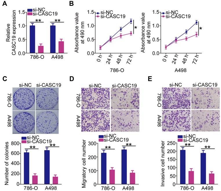
Methods: CASC19 expression was measured by using reverse transcription-quantitative polymerase chain reaction. The effects of CASC19 on ccRCC cell proliferation, colony formation, migration, and invasiveness in vitro, as well as on tumor growth in vivo, were examined by the MTT assay, colony formation assay, cell migration and invasiveness assays, and tumor xenograft in nude nice, respectively.

Results: CASC19 was overexpressed in ccRCC tissues and cell lines. High expression of CASC19 was closely associated with unfavorable clinicopathological parameters and predicted negative clinical outcomes in patients with ccRCC. Knockdown of CASC19 decreased ccRCC cell proliferation, colony formation, migration, and invasiveness, as well as attenuated tumor growth in vivo. Mechanistically, CASC19 functioned as a competing endogenous RNA and upregulated the expression of ETS proto-oncogene 1 (ETS1) through sponging microRNA-532 (miR-532). Furthermore, rescue assays revealed that inhibiting miR-532 or restoring ETS1 expression partially abolished the impacts of CASC19 knockdown on ccRCC cells.

Conclusion: The CASC19/miR-532/ETS1 regulatory pathway is crucial for the malignant manifestations of ccRCC, which makes it an attractive target for potential treatments of ccRCC.

Keywords: MTT assay; cancer; cell migration; invasiveness; knockdown; xenograft.


Conflict of interest statement

The authors declare that they have no competing interests in this work.

Figures

Figure 1
Figure 1
Upregulation of CASC19 in ccRCC tissues and cell lines (A) Relative CASC19 expression detected in 51 pairs of ccRCC and adjacent normal renal tissue samples by using RT-qPCR.(B) CASC19 expression levels determined by RT-qPCR in three ccRCC cell lines (786-O, Caki-1, and A498) and the normal human proximal tubule epithelial cell line HK-2. (C) ccRCC patients with high CASC19 expression had much lower overall survival rate than those with low CASC19 expression (P = 0.038). Statistical significance of differences is indicated as follows: *P <0.05 and **P <0.01.
Figure 2
Figure 2
Attenuation of 786-O and A498 cell proliferation, migration, and invasiveness in vitro by CASC19 knockdown (A) CASC19 levels determined by RT-qPCR in 786-O and A498 cells transfected with si-CASC19 or si-NC. (B, C) The modulatory effect of CASC19 downregulation on the proliferation and colony formation of 786-O and A498 cells determined by the MTT and colony formation assays. (D, E) Migratory and invasive capacities of 786-O and A498 cells measured by the cell migration and invasion assays after CASC19 knockdown. Statistical significance of differences is indicated as follows: *P<0.05 and **P <0.01.
Figure 3
Figure 3
CASC19 functions as a molecular sponge for miR-532 in ccRCC cells. (A) 786-O and A498 cells were fractionated into nuclear and cytosolic fractions. Total RNA of each fraction was isolated and CASC19 expression level was determined by using RT-qPCR. (B) The schematic of miR-532 wild-type (wt) and mutant (mut) targeting sites within CASC19. (C) miR-532 levels in 786-O and A498 cells transfected with miR-532 mimics or miR-NC were measured by RT-qPCR. (D) Luciferase activity of the CASC19-wt or CASC19-mut constructs determined in the presence of miR-532 mimics or miR-NC. (E) 786-O and A498 cell lysates were immunoprecipitated with AGO2 or IgG antibodies. The immunoprecipitated RNA was subjected to RT-qPCR analysis to quantify miR-532 and CASC19 levels. (F) miR-532 expression levels in 51 pairs of ccRCC and adjacent normal renal tissue samples determined by using RT-qPCR. (G) Correlation between miR-532 and CASC19 expression levels in 51 ccRCC tissue samples (r = −0.6180, P < 0.0001). (H) miR-532 expression levels in CASC19-deficient 786-O and A498 cells determined by using RT-qPCR. Statistical significance of differences is indicated as follows: *P < 0.05 and **P < 0.01.
Figure 4
Figure 4
CASC19 directly sponges miR-532 expression and thereby positively regulates ETS1 expression in ccRCC tissues. (A, B) Effects of CASC19 silencing on ETS1 mRNA and protein expression levels in 786-O and A498 cells determined by using RT-qPCR and Western blotting, respectively. (C) Expression of ETS1 mRNA in ccRCC and adjacent normal renal tissues analyzed by using RT-qPCR. (D) Correlation between expression levels of CASC19 and ETS1 mRNA in 51 samples of ccRCC tissues (r = 0.4738, P = 0.0004). (E) Correlation between expression levels of miR-532 and ETS1 mRNA in 51 samples of ccRCC tissues (r = −0.5296, P = 0.0010). (F) miR-532 expression levels in 786-O and A498 cells at 48 h after transfection with the miR-532 inhibitor or NC inhibitor. (G, H) ETS1 mRNA and protein expression levels quantified by RT-qPCR and Western blotting, respectively, in 786-O and A498 cells transfected with si-CASC19 and either the miR-532 inhibitor or NC inhibitor. Statistical significance of differences is indicated as follows: *P <0.05 and **P <0.01.
Figure 5
Figure 5
Inhibition of miR-532 alleviates the inhibitory effects of CASC19 knockdown in 786-O and A498 cells. (AD) Proliferation, colony formation, migration, and invasiveness properties of 786-O and A498 cells after co-transfection with si-CASC19 and either miR-532 inhibitor or NC inhibitor determined in the MTT, colony formation, cell migration and invasion assays. *P <0.05 and **P <0.01.
Figure 6
Figure 6
Restoration of ETS1 expression abrogates the inhibitory effects of CASC19 knockdown in 786-O and A498 cells. (A) ETS1 protein expression in 786-O and A498 cells transfected with pcDNA3.1-ETS1 or pcDNA3.1 measured by Western blot. (BE) Proliferation, colony formation, migration, and invasiveness properties of 786-O and A498 cells after their co-transfection with si-CASC19 and either pcDNA3.1-ETS1 or pcDNA3.1 determined in the MTT, colony formation, cell migration and invasion assays. Statistical significance of differences is indicated as follows: *P <0.05 and **P <0.01.
Figure 7
Figure 7
CASC19 depletion impairs tumor growth of ccRCC cells in vivo. (A) CASC19 expression in A498 cells stably transfected with sh-CASC19 quantified by using RT-qPCR. (B) Weekly measurements of tumor xenograft volume. (C) Representative images of tumor xenografts obtained from ccRCC cells transfected with sh-CASC19 and sh-NC. (D) Tumor xenograft weight determined when the nude mice were euthanized at 4 weeks post-inoculation. (E) miR-532 expression level in tumor xenografts quantified by using RT-qPCR. (F) Western blot analysis of ETS1 protein expression in tumor xenografts. Statistical significance of differences is indicated as follows: *P <0.05 and **P <0.01.

References

    1. Garcia JA, Cowey CL, Godley PA. Renal cell carcinoma. Curr Opin Oncol. 2009;21(3):266–271. doi:10.1097/CCO.0b013e32832a05c8 - DOI - PubMed
    1. Sourbier C, Danilin S, Lindner V, et al. Targeting the nuclear factor-kappaB rescue pathway has promising future in human renal cell carcinoma therapy. Cancer Res. 2007;67(24):11668–11676. doi:10.1158/0008-5472.CAN-07-0632 - DOI - PubMed
    1. Hsieh JJ, Purdue MP, Signoretti S, et al. Renal cell carcinoma. Nature Rev Dis Primers. 2017;3:17009. doi:10.1038/nrdp.2017.9 - DOI - PMC - PubMed
    1. Chow WH, Dong LM, Devesa SS. Epidemiology and risk factors for kidney cancer. Nature Rev Urol. 2010;7(5):245–257. doi:10.1038/nrurol.2010.46 - DOI - PMC - PubMed
    1. Greef B, Eisen T. Medical treatment of renal cancer: new horizons. Br J Cancer. 2016;115(5):505–516. doi:10.1038/bjc.2016.230 - DOI - PMC - PubMed

Publication types