

Mobile Thrombus in the Ascending Aorta
- PMID: 32273925
- PMCID: PMC7140152
- DOI: 10.3400/avd.cr.19-00063
Mobile Thrombus in the Ascending Aorta
Abstract
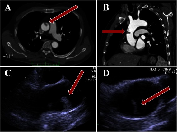
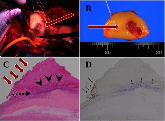
A 65-year-old male who presented with dizziness, dysarthria, and disability of his left hand was admitted to our hospital. Magnetic resonance imaging of the head revealed cerebral infarction and enhanced computed tomography revealed a suspicious thrombus in the ascending aorta. He did not have a coagulation disorder. We performed ascending aortic replacement and removed the thrombus with the aortic wall in order to avoid any recurrences. Here we report the successful treatment of the case from clinical and pathological points of view with some findings.
Keywords: aorta; graft-replacement; thrombus.
Copyright © 2020 Annals of Vascular Diseases.
Conflict of interest statement
Disclosure StatementAll authors have no conflict of interest.
Figures
References
-
- Jaworski L, Fijalkowski M, Rogowski J. Giant thrombus in ascending aorta and aortic arch. J Thorac Cardiovasc Surg 2013; 145: 1668-9. - PubMed
-
- Haida H, Inoue Y, Kawajiri H, et al. Rod-shaped giant floating thrombus in a normal ascending aorta. Eur J Cardiothorac Surg 2015; 47: 582. - PubMed
-
- Fayad ZY, Semaan E, Fahoum B, et al. Aortic mural thrombus in the normal or minimally atherosclerotic aorta. Ann Vasc Surg 2013; 27: 282-90. - PubMed
-
- Shimizu H, Tanibuchi A, Akaishi M, et al. Stroke due to undifferentiated aortic intimal sarcoma with disseminated metastatic lesions. Circulation 2009; 120: e290-2. - PubMed
-
- Rhee MY, Myong NH, Park YB. Primary intimal sarcoma of the aorta: role of transesophageal echocardiography. Circ J 2002; 66: 111-3. - PubMed