

Diagnostic value of echocardiography on detecting the various types of anomalous origin of the left coronary artery from the pulmonary artery
- PMID: 32274098
- PMCID: PMC7139093
- DOI: 10.21037/jtd.2020.01.28
Diagnostic value of echocardiography on detecting the various types of anomalous origin of the left coronary artery from the pulmonary artery
Abstract
Background: To assess the diagnostic value of echocardiography in detecting the various types of anomalous origin of the left coronary artery from the pulmonary artery (ALCAPA).
Methods: A total of 30 patients with an established diagnosis of ALCAPA were retrospectively analyzed, and classified into infant- (n=20) and adult-type (n=10) groups according to the age of symptom manifestation and the mode of presentation. All patients underwent echocardiography examination.
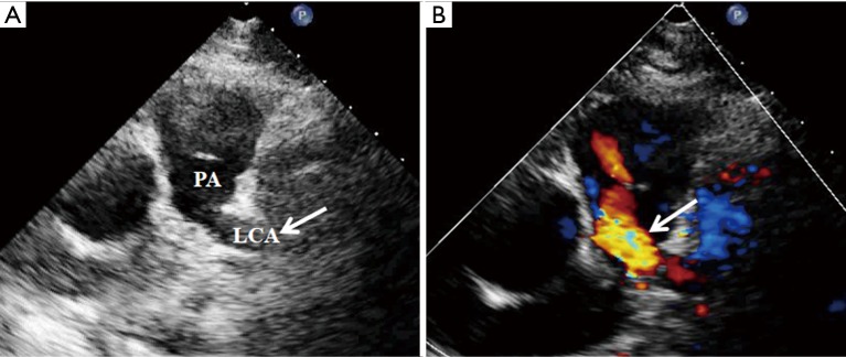
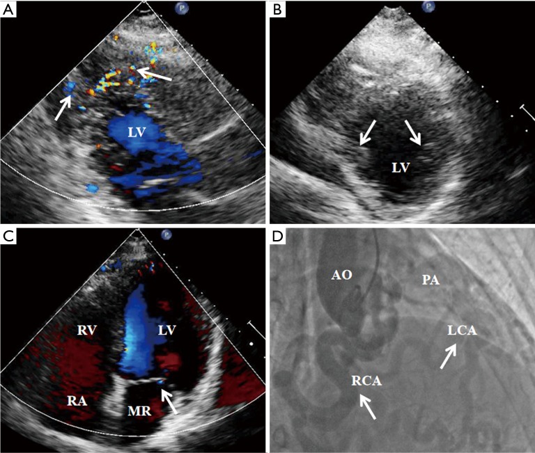
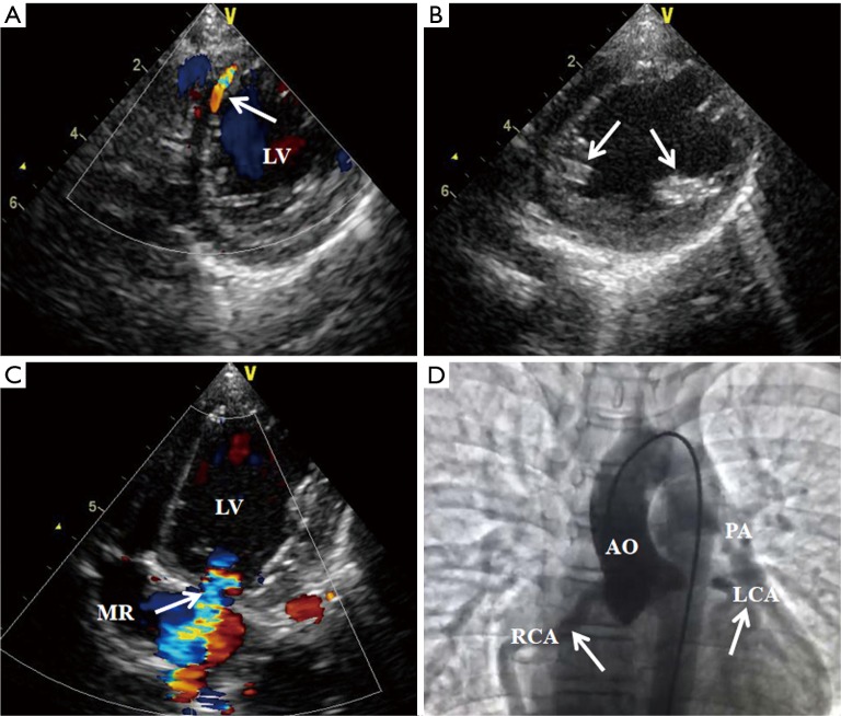
Results: Twenty-four out of thirty patients were diagnosed with ALCAPA by echocardiography. The remaining six cases were confirmed by dual-source computed tomography (DSCT) and angiocardiography, respectively. In the infant-type group, there was negligible or no collateral flow between the right coronary artery (RCA) and the left coronary artery (LCA). Eighteen of these patients had enhanced echogenicity of left ventricular (LV) papillary muscles, different degrees of mitral regurgitation (MR) and the RCA to aortic annulus ratio (RCA/AO) was >0.12. In the adult-type group, all ten patients had RCA dilation and significant development of collateralization from the RCA to the dilated LCA. They all had mild MR and RCA/AO was >0.20. Preoperatively, left ventricular ejection fraction (LVEF) was significantly lower in infant-type group than in adult-type group (46.24%±5.47% vs. 61.43%±6.38%, P<0.01). Cardiac surgery significantly improved post-operative LVEF (60.12%±6.02%, P<0.01 vs. pre-operation) in infant-type group.
Conclusions: Echocardiography plays a pivotal role in detecting ALCAPA. Imaging and clinical features differ significantly between infant- and adult-type cases.
Keywords: Echocardiography; adult; anomalous origin of the left coronary artery from the pulmonary artery (ALCAPA); infant.
2020 Journal of Thoracic Disease. All rights reserved.
Conflict of interest statement
Conflicts of Interest: The authors have no conflicts of interest to declare.
Figures
Similar articles
-
Two congenital coronary abnormalities affecting heart function: anomalous origin of the left coronary artery from the pulmonary artery and congenital left main coronary artery atresia.Chin Med J (Engl). 2014;127(21):3724-31. Chin Med J (Engl). 2014. PMID: 25382327
-
Transthoracic echocardiography features of adult-type anomalous left coronary artery from the pulmonary artery before and after surgery: highlights from observational study in a single center of China.Int J Cardiovasc Imaging. 2020 Aug;36(8):1477-1487. doi: 10.1007/s10554-020-01857-x. Epub 2020 Apr 23. Int J Cardiovasc Imaging. 2020. PMID: 32328871
-
[Analysis on missed diagnosis or misdiagnosis of anomalous origin of left coronary artery from pulmonary artery by echocardiography from one single medical center].Zhonghua Xin Xue Guan Bing Za Zhi. 2023 May 24;51(5):481-489. doi: 10.3760/cma.j.cn112148-20220712-00541. Zhonghua Xin Xue Guan Bing Za Zhi. 2023. PMID: 37198119 Chinese.
-
Myocardial function in patients with anomalous left coronary artery from the pulmonary artery syndrome: A long-term speckle tracking echocardiographic study.PLoS One. 2019 Oct 15;14(10):e0223227. doi: 10.1371/journal.pone.0223227. eCollection 2019. PLoS One. 2019. PMID: 31613933 Free PMC article.
-
Anomalous Left Coronary Artery from the Pulmonary Artery: How to Diagnose and Treat.J Pers Med. 2023 Oct 31;13(11):1561. doi: 10.3390/jpm13111561. J Pers Med. 2023. PMID: 38003878 Free PMC article. Review.
Cited by
-
Anatomical Variants of the Origin of the Coronary Arteries: A Systematic Review and Meta-Analysis of Prevalence.Diagnostics (Basel). 2024 Jul 8;14(13):1458. doi: 10.3390/diagnostics14131458. Diagnostics (Basel). 2024. PMID: 39001347 Free PMC article. Review.
-
Anomalous Origin of the Coronary Artery from the Pulmonary Artery in Children and Adults: A Pictorial Review of Cardiac Imaging Findings.Korean J Radiol. 2021 Sep;22(9):1441-1450. doi: 10.3348/kjr.2021.0034. Epub 2021 May 20. Korean J Radiol. 2021. PMID: 34047508 Free PMC article. Review.
-
Angina Pectoris as a Manifestation of ALCAPA Syndrome in a 20-Year-Old Female: A Case Report and Review of Literature.Eur J Case Rep Intern Med. 2023 Jun 26;10(7):003962. doi: 10.12890/2023_003962. eCollection 2023. Eur J Case Rep Intern Med. 2023. PMID: 37455696 Free PMC article.
-
Anomalous left coronary artery from the pulmonary artery in a symptomatic adult: a case report.Ann Med Surg (Lond). 2024 Aug 8;86(9):5622-5626. doi: 10.1097/MS9.0000000000002451. eCollection 2024 Sep. Ann Med Surg (Lond). 2024. PMID: 39239055 Free PMC article.
-
Clinical Characteristics of Congenital Atresia of the Left Main Coronary Artery in 12 Children.Front Pediatr. 2022 Apr 29;10:866010. doi: 10.3389/fped.2022.866010. eCollection 2022. Front Pediatr. 2022. PMID: 35573961 Free PMC article.
References
LinkOut - more resources
Full Text Sources
Research Materials