. 2020 Mar;12(3):383-393.
doi: 10.21037/jtd.2020.01.34.

Pulmonary veins variations with potential impact in thoracic surgery: a computed-tomography-based atlas

Affiliations

Pulmonary veins variations with potential impact in thoracic surgery: a computed-tomography-based atlas

Mateusz Polaczek et al. J Thorac Dis. 2020 Mar.

Abstract

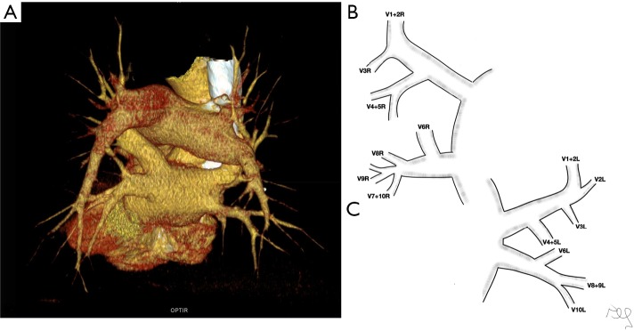
Background: Pulmonary veins (PVs) are important during segmentectomy. Many case reports prove that they may be the source of bleeding during surgery, especially when anatomical variants are present. We decided to describe venous variations and prepare a computed tomography based atlas of our observations.

Methods: The study was conducted using 135 chest computed tomography studies with intra venous iodine contrast injection. The study population contained 86 females and 49 males, mean age was 60. Thirteen people had atrial fibrillation. Images were analysed using radiological workstation.

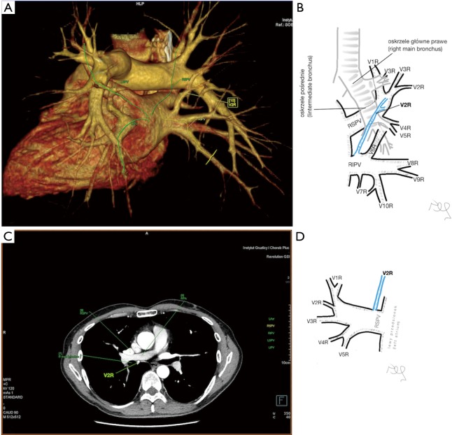
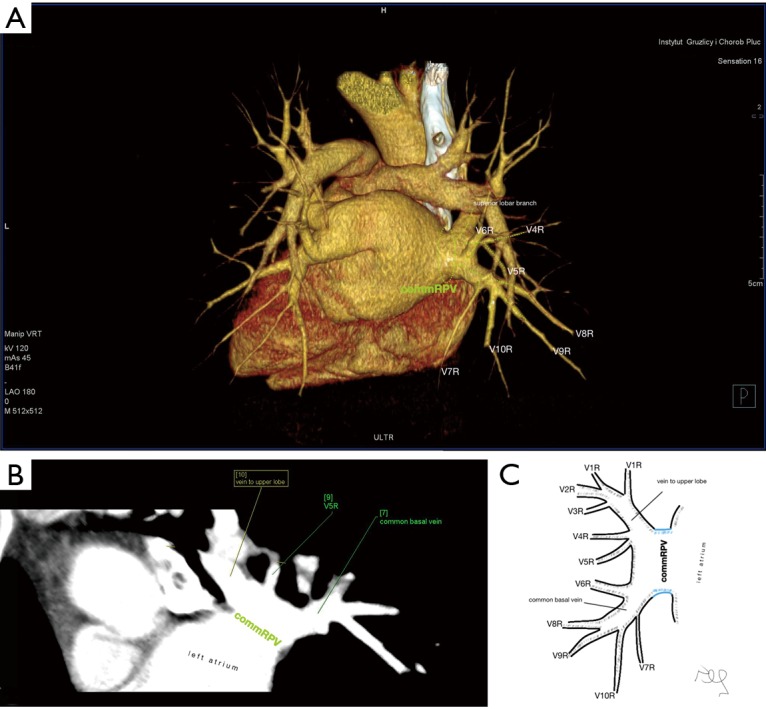
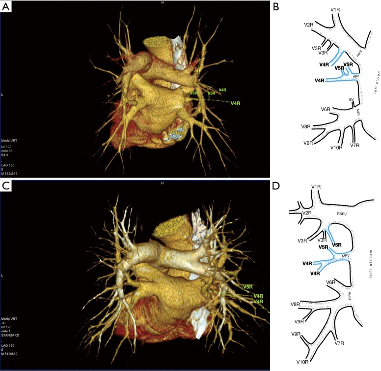
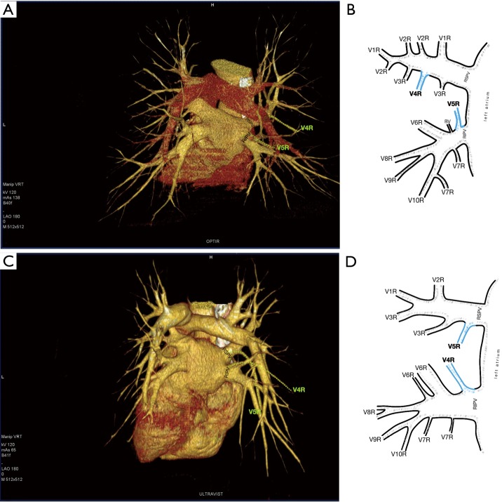
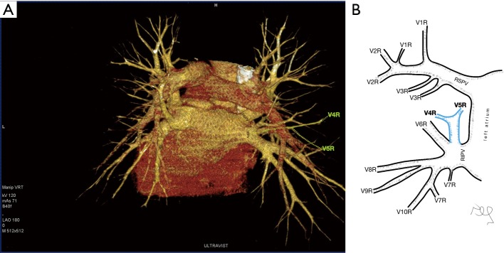
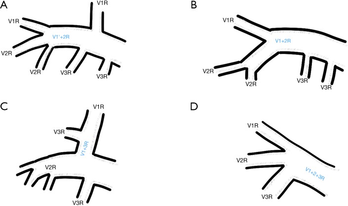
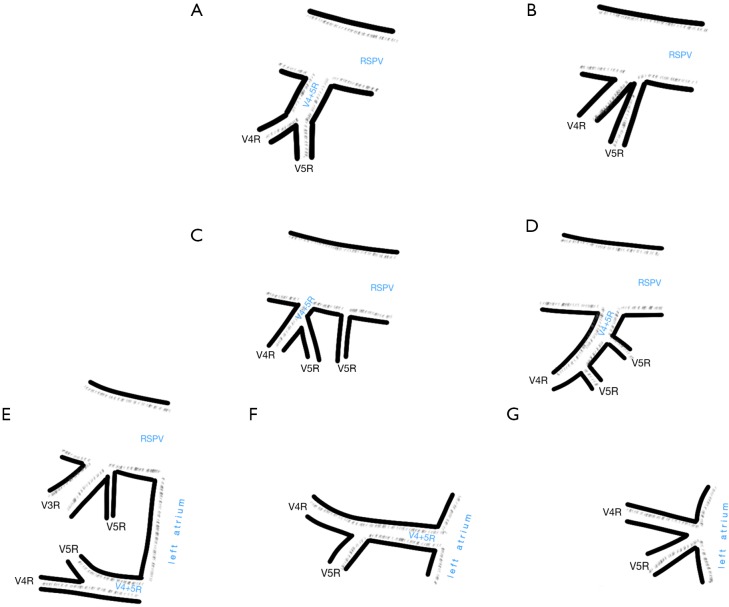
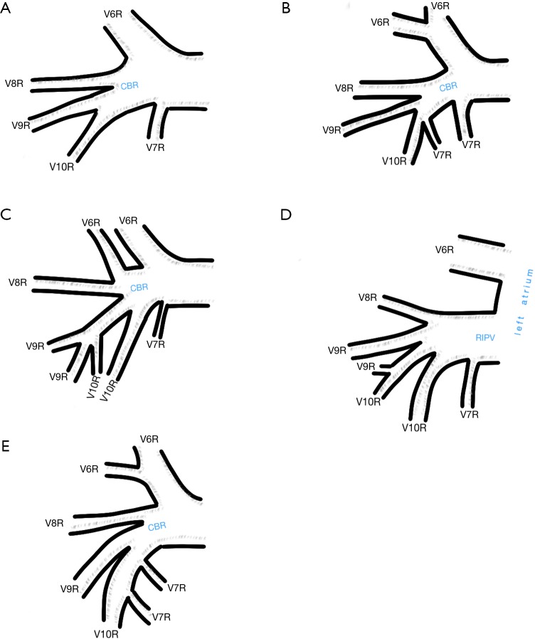
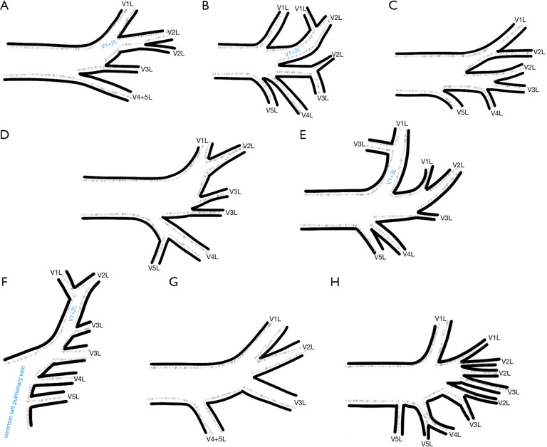
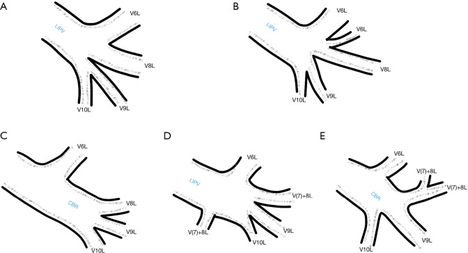
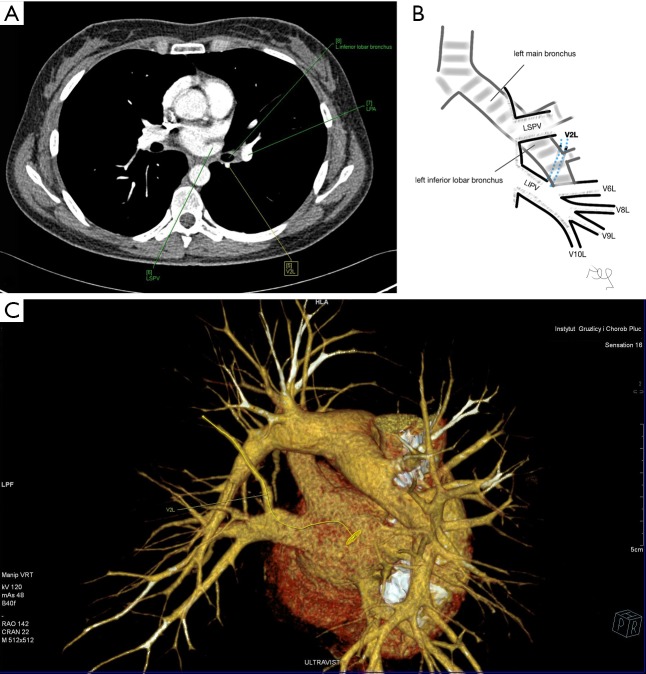
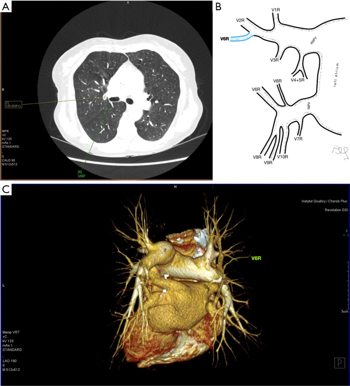
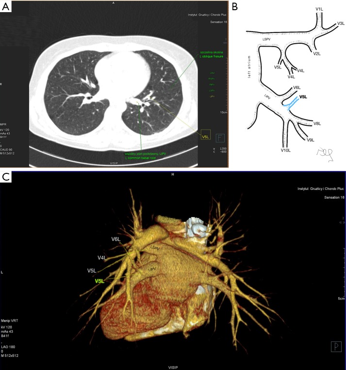
Results: The variations were divided into three categories: atypical topography of the PV, atypical venous outflow to the left atrium (LA), atypical venous vascularization of the lung bronchopulmonary segment. Retrobronchial course of the vein of the posterior segment of the right upper lobe was observed in 8.15%. The most common variant of atrial venous outflow was the direct outflow of the middle lobe vein, observed in 25.19% of cases and the long common trunk of left PVs in 11.11%. The split drainage from the middle lobe into the right superior pulmonary vein (RSPV) and the right inferior pulmonary vein (RIPV) was observed in 9.63% as the full drainage into the RIPV in 2.96%.

Conclusions: Long common trunk of left PVs and numerous variants of venous vascularisation of the middle lobe are the variations that may pose potential problems during thoracic surgeries. The frequency is high enough to justify the routine assessment of pulmonary vessels with computed tomography before surgery.

Keywords: Pulmonary veins (PVs); anatomy; computed tomography (CT); vascular complications.


Conflict of interest statement

Conflicts of Interest: The authors have no conflicts of interest to declare.

Figures

Figure 1
Figure 1
Variant topography of the vein of the posterior segment of the right upper lobe (V2R). V2R runs posteriorly to the intermediate bronchus and empties into the trunk of the right inferior pulmonary vein (RIPV): (A) CT with VRT, posterior view; (B) schematic posterior left oblique view. V2R runs medially to the intermediate bronchus and crosses posteriorly the trunk of the right pulmonary artery; (C) CT, axial plane, mediastinal window; (D) schematic anterior view. CT, computed tomography; VRT, volume rendering technique.
Figure 2
Figure 2
The vein of the apical segment of the right lower lobe (V6R), runs posteriorly to the intermediate bronchus and outflows directly to the left atrium. (A) CT with VRT, posterior view; (B) schematic posterior left oblique view. CT, computed tomography; VRT, volume rendering technique.
Figure 3
Figure 3
The vein of the superior segment of the upper right lobe (V1R) runs medially and places between the superior vena cava (SVC) and the right pulmonary artery (RPA). (A) CT with VRT, left posterior oblique view; (B) schematic representation of V1R with relation to RPA, SVC, anterior view; CT, computed tomography; VRT, volume rendering technique.
Figure 4
Figure 4
Common trunk of left pulmonary veins (commLPV) composed of multiple branches to the upper lobe segments and lower lobe trunk. (A) CT with VRT, posterior view; (B) CT with CPR; (C) schematic anterior view. CT, computed tomography; VRT, volume rendering technique; CPR, curved planar reformations.
Figure 5
Figure 5
Common trunk of right pulmonary veins (commRPV) composed of two short branches. (A) CT with VRT, posterior view; (B) CT with CPR; (C) schematic anterior view. CT, computed tomography; VRT, volume rendering technique; CPR, curved planar reformations.
Figure 6
Figure 6
Split drainage from the middle lobe. Direct outflow of the middle lobe vein (MPV) to the left atrium (LA) and accessory vein from the lateral segment of the middle lobe (V4R) running into the right superior pulmonary vein (RSPV): (A) CT with VRT, posterior view; (B) schematic anterior view. Direct outflow of MPV to LA and accessory vein from the medial segment of the middle lobe (V5R) running into the vein of the anterior segment of the upper right lobe (V3R): (C) CT with VRT, posterior view; (D) schematic anterior view. CT, computed tomography; VRT, volume rendering technique.
Figure 7
Figure 7
Split drainage from the middle lobe. V4R flows into RSPV and V5R flows into the right inferior pulmonary veins (RIPV): (A) CT with VRT, posterior view; (B) schematic anterior view. V5R runs into RSPV and V4R outflows into RIPV: (C) CT with VRT, posterior view; (D) schematic anterior view. RSPV, right superior pulmonary vein; CT, computed tomography; VRT, volume rendering technique.
Figure 8
Figure 8
Full drainage of the middle lobe to the right inferior pulmonary vein. (A) CT with VRT, posterior view; (B) schematic anterior view. CT, computed tomography; VRT, volume rendering technique.
Figure S1
Figure S1
Normal variant of the segmental vascularisation of the lungs. (A) CT with VRT, posterior view; (B) right pulmonary veins; (C) left pulmonary veins. CT, computed tomography; VRT, volume rendering technique.
Figure S2
Figure S2
Most common segmental veins of the right upper lobe.
Figure S3
Figure S3
Most common segmental veins of the middle lobe. RSPV, right superior pulmonary vein.
Figure S4
Figure S4
Most common segmental veins of the right lower lobe. CBR, common basal root; RIPV, right inferior pulmonary vein.
Figure S5
Figure S5
Most common segmental veins of the left upper lobe.
Figure S6
Figure S6
Most common segmental veins of the left lower lobe. CBR, common basal root; LIPV, left inferior pulmonary vein.
Figure S7
Figure S7
Retrobronchial course of the vein of the posterior segment of the left upper lobe (V2L) with outflow to the left inferior pulmonary vein (LIPV) close to V6L. (A) CT, axial plane, mediastinal window; (B) schematic anterior view; (C) CT with VRT, right posterior oblique view. CT, computed tomography; VRT, volume rendering technique.
Figure S8
Figure S8
Accessory vein of the apical segment of the right lower lobe (V6R) connects with the vein of the posterior segment of the right upper lobe (V2R). (A) CT, axial plane, lung window; (B) schematic anterior view; (C) CT with VRT, posterior view. CT, computed tomography; VRT, volume rendering technique.
Figure S9
Figure S9
Partial outflow from the lingua of the upper left lobe to the left inferior pulmonary vein (LIPV): the vein of the inferior lingual segment (V5L) drains into LIPV. (A) CT, axial plane, lung window; (B) schematic anterior view; (C) CT with VRT, posterior view. CT, computed tomography; VRT, volume rendering technique.

References

    1. Pearson FG, Cooper JD, Deslauriers J, et al. editors. Troracic Surgery. Second Edition. Philadelphia, Pensylvania: Churchill Livingstone, 2002:981-90.
    1. Gonzalez-Rivas D, Yang Y, Ng C. Advances in Uniportal Video-Assisted Thoracoscopic Surgery. Thorac Surg Clin 2016;26:187-201. 10.1016/j.thorsurg.2015.12.007 - DOI - PubMed
    1. Oizumi H, Kato H, Endoh M, et al. Techniques to define segmental anatomy during segmentectomy. Ann Cardiothorac Surg 2014;3:170. - PMC - PubMed
    1. Swanson SJ. Segmentectomy for Lung Cancer. Semin Thorac Cardiovasc Surg 2010;22:244-9. 10.1053/j.semtcvs.2010.10.014 - DOI - PubMed
    1. Iwano S, Yokoi K, Taniguchi T, et al. Planning of segmentectomy using three-dimensional computed tomography angiography with a virtual safety margin: Technique and initial experience. Lung Cancer 2013;81:410-5. 10.1016/j.lungcan.2013.06.001 - DOI - PubMed