

Review
doi: 10.1016/j.ekir.2019.12.007.
eCollection 2020 Apr.
Polycystins, ADPKD, and Cardiovascular Disease
Affiliations
- PMID: 32274448
- PMCID: PMC7136326
- DOI: 10.1016/j.ekir.2019.12.007
Item in Clipboard
Review
Polycystins, ADPKD, and Cardiovascular Disease
Kidney Int Rep.
.
Abstract
Cardiovascular disorders are the most common cause of mortality in autosomal dominant polycystic kidney disease (ADPKD). This review considers recent clinical and basic science studies that address the contributing factors of cardiovascular dysfunction in ADPKD. In particular, attention is placed on how dysfunction of the polycystin proteins located in the cardiovascular system contributes to extrarenal manifestations of ADPKD.
Keywords: calcium signaling; cardiovascular dysfunction; heart failure; polycystic kidney disease.
© 2019 International Society of Nephrology. Published by Elsevier Inc.
Figures
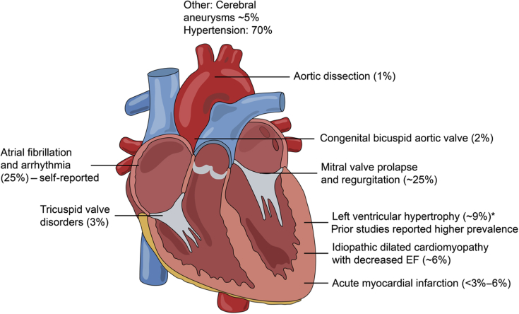
Spectrum of cardiovascular disorders reported in autosomal dominant polycystic kidney disease. Note that prevalence is based on more recent clinical studies (i.e., the past decade, in which blood pressure control has been administered). Please refer to the text for references. EF, ejection fraction.
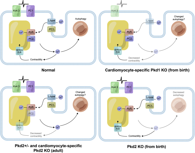
Current understanding of the location and functional roles of polycystin (PC)1 and PC2 in cardiomyocytes. Normal: PC1 appears to be on the plasma membrane in cardiomyocytes and has been shown to interact with the potassium (Kv) channel 4.3 and regulate the expression of the L-type calcium channel (VDCC). PC2 has been shown to interact with the intracellular release channel ryanodine receptor (RyR). Arrows point to the movement of calcium in a cardiomyocyte contributing to contractility and autophagy. It is unclear if PC1 and PC2 interact directly with each other in cardiomyocytes, as demonstrated in the renal epithelial cells. PC1 knockout (KO) models: KO of PC1 from birth leads to decreased L-type VDCC; decreased Kv4.3 expression, reduced action potential duration, decreased calcium release, and decreased contractility. Autophagy in cardiomyocytes has not been addressed yet in the Pkd1 KO mouse. PC2 models: Heterozygous and adult induced PC2 KO causes increased released calcium and, paradoxically, decreased contractility due to desensitization of the myofilament to calcium. The calcium uptake mechanism into the sarcoplasmic reticulum via SERCA and phospholamban is less active. From-birth KO Pkd2: From-birth KO of cardiac PC2 causes decreased released calcium and decreased autophagy. SERCA, sarco/endoplasmic reticulum Ca2+-ATPase.
References
Publication types
LinkOut - more resources
Full Text Sources
