. 2021 Mar;31(1):245-256.
doi: 10.1007/s00062-020-00879-1. Epub 2020 Apr 9.

Presurgical Localization of the Primary Sensorimotor Cortex in Gliomas : When is Resting State FMRI Beneficial and Sufficient?

Affiliations

Presurgical Localization of the Primary Sensorimotor Cortex in Gliomas : When is Resting State FMRI Beneficial and Sufficient?

Natalie L Voets et al. Clin Neuroradiol. 2021 Mar.

Abstract

Purpose: Functional magnetic resonance imaging (fMRI) has an established role in neurosurgical planning; however, ambiguity surrounds the comparative value of resting and task-based fMRI relative to anatomical localization of the sensorimotor cortex. This study was carried out to determine: 1) how often fMRI adds to prediction of motor risks beyond expert neuroradiological review, 2) success rates of presurgical resting and task-based sensorimotor mapping, and 3) the impact of accelerated resting fMRI acquisitions on network detectability.

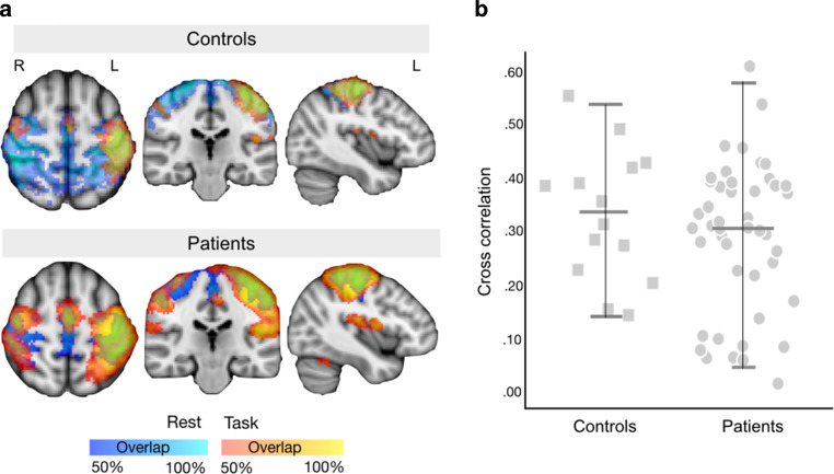
Methods: Data were collected at 2 centers from 71 patients with a primary brain tumor (31 women; mean age 41.9 ± 13.9 years) and 14 healthy individuals (6 women; mean age 37.9 ± 12.7 years). Preoperative 3T MRI included anatomical scans and resting fMRI using unaccelerated (TR = 3.5 s), intermediate (TR = 1.56 s) or high temporal resolution (TR = 0.72 s) sequences. Task fMRI finger tapping data were acquired in 45 patients. Group differences in fMRI reproducibility, spatial overlap and success frequencies were assessed with t‑tests and χ2-tests.

Results: Radiological review identified the central sulcus in 98.6% (70/71) patients. Task-fMRI succeeded in 100% (45/45). Resting fMRI failed to identify a sensorimotor network in up to 10 patients; it succeeded in 97.9% (47/48) of accelerated fMRIs, compared to only 60.9% (14/23) of unaccelerated fMRIs ([Formula: see text](2) = 17.84, p < 0.001). Of the patients 12 experienced postoperative deterioration, largely predicted by anatomical proximity to the central sulcus.

Conclusion: The use of fMRI in patients with residual or intact presurgical motor function added value to uncertain anatomical localization in just a single peri-Rolandic glioma case. Resting fMRI showed high correspondence to task localization when acquired with accelerated sequences but offered limited success at standard acquisitions.

Keywords: ICA; Sensorimotor; Surgery; Task fMRI; Tumor.


Conflict of interest statement

N.L. Voets, P. Plaha, O.P. Jones, P. Pretorius and A. Bartsch declare that they have no competing interests.

Figures

Fig. 1
Fig. 1
Anatomical landmarks identifying the central sulcus. Two consultant neuroradiologists independently identified the central sulci (CS) in the 71 participants’ T1-weighted anatomical scan. a Template MNI152 brain illustrating the combination of four well-established landmarks (red lines) used to locate the CS (blue line): the inverted omega which can be shallow or doubled (see Fig. 3); the inverted T sign; the “pli de passage fronto-pariétal supérieur”; and the termination of precentral gyrus behind pars opercularis. The lowest extension of the precentral gyrus posteriorly turns into the subcentral gyrus (not marked), which is also helpful for orientation. b Application of these landmarks to localize the central sulcus in a patient (Case 15 in Table 4) with severe effacement of the CS due to tumor infiltration and mass effect. Here, the location of the CS (blue line) was not unambiguous but determined on consensus re-review to course through the body of the tumor (outlined in red on the 3D brain rendering)
Fig. 2
Fig. 2
Task and resting fMRI-derived (sensori)motor maps. a Overlap heat maps in controls (n = 14) and patients (n = 45 task, n = 71 rest) for task (red-yellow) and resting (blue) fMRI overlaid onto the template MNI152 brain. Only those voxels shared by at least 50% of each group are shown. b Overlap between task and resting maps was computed using spatial cross-correlation. There was no statistical difference in the amount of overlap between task and resting fMRI maps between healthy controls and glioma patients (independent samples t‑test, t = 0.96, p = 0.35)
Fig. 3
Fig. 3
Lack of apparent functional reorganization in individual tfMRI maps. Single subject tfMRI results during finger tapping were visualized to examine evidence for potential functional reorganization in 6 patients with a tumor directly encroaching upon or distorting the peri-Rolandic cortex. All patients had normal muscle power (5/5 on the MRC scale). In each case, the task activation localized to the anatomically expected region on the motor homunculus in the hemisphere contralateral to the hand used to perform the task, and ipsilateral to the tumor. Yellow stars mark the glioma in each case
Fig. 4
Fig. 4
Visual identification of sensorimotor components in individuals’ resting fMRI. Independent Component Analysis (ICA) decomposition of the resting fMRI time series in two representative patients. a In some patients (typically with basic clinical rsfMRI acquisitions of low temporal resolution) a single extensive, bilateral sensorimotor network was identified. Visual network identification relied upon 3 criteria: spatial correspondence with the central sulcus (top left), a biologically plausible time-course (top right) and a predominantly low-frequency (0.01–0.05 Hz) power spectrum (bottom right). b In another patient, scanned at a high temporal resolution rsfMRI, multiple spatially segregated sensorimotor resting networks were identified, which co-localized with the expected functional divisions of the foot/leg (green), hand/arm (red/yellow; split by hemisphere) and mouth/face (blue) areas along the central sulcus

Similar articles

Cited by

References

    1. Kim YH, Kim CH, Kim JS, Lee SK, Han JH, Kim CY, et al. Topographical risk factor analysis of new neurological deficits following precentral gyrus resection. Neurosurgery. 2015;76(6):714–720. doi: 10.1227/NEU.0000000000000712. - DOI - PubMed
    1. Penfield W, Boldrey E. Somatic motor and sensory representation in the cerebral cortex of man as studied by electrical stimulation. Brain. 1937;60:389–443. doi: 10.1093/brain/60.4.389. - DOI
    1. Rademacher J, Burgel U, Geyer S, Schormann T, Schleicher A, Freund HJ, et al. Variability and asymmetry in the human precentral motor system. A cytoarchitectonic and myeloarchitectonic brain mapping study. Brain. 2001;124(11):2232–2258. doi: 10.1093/brain/124.11.2232. - DOI - PubMed
    1. Nii Y, Uematsu S, Lesser RP, Gordon B. Does the central sulcus divide motor and sensory functions? Cortical mapping of human hand areas as revealed by electrical stimulation through subdural grid electrodes. Neurology. 1996;46(2):360–367. doi: 10.1212/WNL.46.2.360. - DOI - PubMed
    1. Wengenroth M, Blatow M, Guenther J, Akbar M, Tronnier VM, Stippich C. Diagnostic benefits of presurgical fMRI in patients with brain tumours in the primary sensorimotor cortex. Eur Radiol. 2011;21(7):1517–1525. doi: 10.1007/s00330-011-2067-9. - DOI - PMC - PubMed